
Guide to Biplane Labs: Advanced Neurovascular Technology Explained
Discover how biplane labs revolutionise neurovascular care with dual-angle imaging, enabling faster, safer, and more precise treatment for stroke and brain aneurysms.

Written by Dr. Md Yusuf Shareef
Reviewed by Dr. Dhankecha Mayank Dineshbhai MBBS
Last updated on 13th Jan, 2026

Introduction
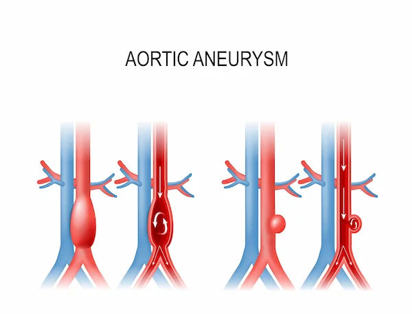
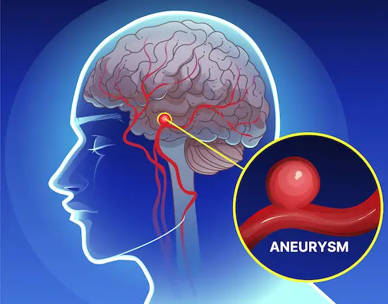
The human brain, with its intricate web of vessels, is one of the body's most delicate and vital organs. When something goes wrong—like a clot blocking blood flow (ischaemic stroke) or a weak spot bulging in an artery (aneurysm)—doctors need to act with incredible speed and accuracy. Enter the biplane lab. Far more advanced than a standard X-ray or CT scanner, a biplane lab is a specialised suite equipped with sophisticated imaging technology that allows physicians to see inside the brain's vasculature in real-time, in three dimensions. This capability is transformative. It enables minimally invasive procedures where doctors can thread tiny instruments through blood vessels to fix problems from within, avoiding risky open-brain surgery. Understanding this technology can provide immense peace of mind, knowing that tools of such precision are available for diagnosing and treating complex neurovascular conditions. This article will serve as your comprehensive guide to this amazing medical advancement.
What is a Biplane Lab? Beyond a Simple X-Ray
At its core, a biplane lab is an angiography suite equipped with a powerful imaging system. The "biplane" name comes from its defining feature: two X-ray imaging systems mounted on rotating arms (C-arms) that move independently around the patient.
The Core Concept: Two Cameras are Better Than One
Imagine trying to pinpoint the exact location of a flying bird with a single photograph. You might get a general idea, but you'd lack depth. Now, imagine using two cameras at different angles simultaneously. This stereo vision gives you a precise, three-dimensional location. This is the fundamental principle of biplane imaging. One X-ray source takes images from the front (anteroposterior view), while the other takes images from the side (lateral view). By combining these two views in real-time, interventional neurologists and radiologists get a dynamic, 3D-like understanding of the complex vascular structures in the brain, all without making a single incision.
A Brief History: From Single-Plane to Biplane Imaging
Before biplane systems became the gold standard for complex interventions, doctors relied on single-plane systems. These had only one X-ray source and detector. To get two different views, the C-arm had to be physically rotated between shots, a process that was time-consuming and could miss crucial, fleeting details during a dynamic procedure. The advent of biplane technology was a monumental leap forward, allowing for simultaneous imaging that drastically reduces procedure time and enhances accuracy, which is critical when every minute counts during a stroke.
Consult a Specialist for the best advice
Why Biplane Technology is a Game-Changer for Neurovascular Care
The advantages of using a biplane lab extend far beyond just taking pictures from two angles. This technology directly translates to better, safer, and faster care for patients.
Unmatched Speed in Stroke Treatment
In an ischaemic stroke, "time is brain." Every minute blood flow is restricted, more brain cells die. Biplane imaging accelerates the life-saving procedure known as mechanical thrombectomy. With real-time, dual-view guidance, a doctor can navigate a catheter to the clot, deploy a stent retriever, and remove the obstruction much more efficiently than with a single-plane system. Studies have shown that reducing procedure time by even 15-20 minutes can significantly improve a patient's long-term recovery prospects.
Pinpoint Accuracy for Delicate Brain Vessels
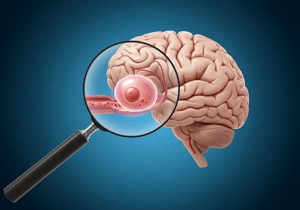
Treating a brain aneurysm is like defusing a bomb; precision is everything. When placing coils or stents inside a tiny aneurysm sac, doctors need to see exactly how the devices are deploying. The dual perspectives of a biplane system provide this spatial awareness, ensuring that the devices are positioned perfectly to block the aneurysm from the inside without compromising the healthy parent artery. This pinpoint accuracy is crucial for preventing ruptures and ensuring the long-term success of the treatment.
Enhanced Safety: Lower Radiation and Contrast Dose
Because a biplane system can capture two views at once, the need to repeatedly take images from different angles is reduced. This means the patient is exposed to less overall radiation. Furthermore, the need for contrast dye (injected to make blood vessels visible) is also minimised. This enhanced safety profile is a significant benefit, especially for patients who may require multiple procedures over time.
Inside the Biplane Lab: Key Technologies at Work
A modern biplane lab is a symphony of integrated technologies. Here’s a breakdown of the key components that make it so effective.
3D Rotational Angiography: Creating a Roadmap of the Brain
This is a standout feature of advanced biplane labs. The C-arm rotates around the patient's head while taking a rapid series of X-rays. A powerful computer then reconstructs these images into a high-resolution, rotatable 3D model of the blood vessels. This gives the physician a detailed "roadmap" before starting the procedure, allowing for better planning and device selection.
Flat Panel Detectors: The Key to Crystal-Clear Images
Modern systems use digital flat panel detectors instead of older image intensifiers. These detectors provide superior image quality with higher contrast and resolution, even at lower radiation doses. This clarity is essential for visualising tiny wires, stents, and coils within the delicate vessels of the brain.
Advanced Software: Real-Time Guidance and Analysis
The hardware is powered by sophisticated software. Features like "road-mapping" allow the doctor to superimpose a live X-ray image over the pre-acquired 3D model, creating a GPS-like guidance system for navigating catheters. Other software can measure blood flow velocity or subtract bone structures from the image to get an unobstructed view of the vessels—a process known as digital subtraction angiography (DSA).
Common Procedures Performed in a Biplane Lab
The precision of biplane technology makes it indispensable for several minimally invasive neurovascular procedures.
Mechanical Thrombectomy for Ischaemic Stroke
This is the primary procedure for large vessel occlusion strokes. A catheter is guided from an artery in the groin or wrist up into the blocked brain artery. A stent retriever is then used to capture and remove the clot, restoring blood flow almost instantly.
Coiling and Stenting for Brain Aneurysms
For a brain aneurysm, a microcatheter is navigated into the aneurysm sac. Platinum coils, which are softer than a human hair, are then packed into the sac to promote clotting and seal it off. In some cases, a stent may be placed in the main artery to act as a scaffold, keeping the coils in place.
Embolisation for Arteriovenous Malformations (AVMs)
An AVM is a tangled web of abnormal blood vessels. Embolisation involves injecting a special glue or particles through a catheter to block off the abnormal vessels, reducing the risk of bleeding. This is often done before surgical removal to make the operation safer. If you or a family member is diagnosed with a complex condition like an AVM, it's crucial to consult a specialist. You can connect with leading neurosurgeons and neurologists through platforms like Apollo24|7 for expert second opinions.
What Patients Can Expect During a Biplane Procedure
If you are scheduled for a procedure in a biplane lab, here’s a general idea of what to expect. You will be given sedation or general anesthesia to ensure you are comfortable and still. The team will sterilise a small area, usually on your groin or wrist, and administer local anesthesia. The doctor will make a tiny incision to access an artery and insert a thin catheter. You will not feel the catheter as it moves through your vessels. The biplane system will move around you, taking images to guide the doctor. The procedure can take anywhere from one to several hours, depending on its complexity. Afterward, you will need to lie flat for a few hours to prevent bleeding from the access site.
The Future is Here: Emerging Trends in Neurovascular Imaging
The evolution of biplane lab technology continues. Integration with artificial intelligence (AI) is the next frontier. AI algorithms can help automate measurements, highlight potential problem areas, and even predict how a device like a stent will behave once deployed. Furthermore, the fusion of biplane angiography with pre-operative MRI or CT scans creates an even more comprehensive navigational map. These advancements promise to make procedures faster, safer, and even more successful.
Conclusion
The development of the biplane lab represents a monumental achievement in medical technology. It has transformed the treatment landscape for neurovascular diseases, offering hope and improved outcomes where options were once limited. By providing physicians with an unparalleled view inside the living brain, this advanced technology enables interventions of breathtaking precision. For patients and families facing the daunting prospect of a stroke or aneurysm, understanding that such sophisticated tools are available can be a great source of comfort. The continued innovation in this field ensures that care will only become more effective and accessible in the years to come. If you have concerns about neurovascular health, always seek timely medical advice. For non-emergency consultations, you can easily book a physical visit or an online appointment with a neurologist through Apollo24|7 to discuss your symptoms and risk factors.
Consult a Specialist for the best advice
Consult a Specialist for the best advice

Dr. Aditendraditya Singh Bhati
Neurosurgeon
22 Years • MBBS(2004), DNB Neurosurgery(2014); MNAMS; Fellow Skull Base Endoscopy (Italy), Fellow Extended Skull Base ( Weill Cornell, USA), Fellow ZAP-X Radiosurgery. Member of American Association of Neurological Surgeons
Delhi
Apollo Hospitals Indraprastha, Delhi
(125+ Patients)

Dr. Ganeshgouda Majigoudra
Neurologist
10 Years • MBBS, MD ( GENERAL MEDICINE) DM (NEUROLOGY)
Bengaluru
Apollo Clinic, JP nagar, Bengaluru

Dr. E Prabhakar Sastry
General Physician/ Internal Medicine Specialist
40 Years • MD(Internal Medicine)
Manikonda Jagir
Apollo Clinic, Manikonda, Manikonda Jagir
(200+ Patients)

Dr. Jaidev S
Neurosurgeon
10 Years • MBBS, MS ( Genera Surgery), MCH Neurolosurgery
Bengaluru
Apollo Clinic, Indiranagar, Bengaluru

Dr. Rajendran S
Neurologist
30 Years • MD, DM (Neuro), MRCP (Irel&), MSc (Neuro)
Chennai
Apollo Hospitals Greams Road, Chennai
(175+ Patients)
Consult a Specialist for the best advice
Dr. Aditendraditya Singh Bhati
Neurosurgeon
22 Years • MBBS(2004), DNB Neurosurgery(2014); MNAMS; Fellow Skull Base Endoscopy (Italy), Fellow Extended Skull Base ( Weill Cornell, USA), Fellow ZAP-X Radiosurgery. Member of American Association of Neurological Surgeons
Delhi
Apollo Hospitals Indraprastha, Delhi
(125+ Patients)
Dr. Ganeshgouda Majigoudra
Neurologist
10 Years • MBBS, MD ( GENERAL MEDICINE) DM (NEUROLOGY)
Bengaluru
Apollo Clinic, JP nagar, Bengaluru
Dr. E Prabhakar Sastry
General Physician/ Internal Medicine Specialist
40 Years • MD(Internal Medicine)
Manikonda Jagir
Apollo Clinic, Manikonda, Manikonda Jagir
(200+ Patients)
Dr. Jaidev S
Neurosurgeon
10 Years • MBBS, MS ( Genera Surgery), MCH Neurolosurgery
Bengaluru
Apollo Clinic, Indiranagar, Bengaluru
Dr. Rajendran S
Neurologist
30 Years • MD, DM (Neuro), MRCP (Irel&), MSc (Neuro)
Chennai
Apollo Hospitals Greams Road, Chennai
(175+ Patients)
More articles from Aneurysm


Frequently Asked Questions
1. Is a procedure in a biplane lab considered surgery?
No, it's considered a minimally invasive endovascular procedure. Instead of a large incision, doctors access the blood vessels through a tiny puncture, usually in the groin or wrist, leading to faster recovery times and fewer risks than traditional open surgery.
2. How does biplane imaging differ from a CT or MRI scan?
A CT or MRI provides a static 'snapshot' of the brain's anatomy. Biplane angiography provides a dynamic, real-time 'movie' of the blood flowing through the vessels. This live feedback is essential for guiding instruments during a procedure.
3. What are the risks of a cerebral angiogram in a biplane lab?
While very safe, risks include bleeding or infection at the puncture site, an allergic reaction to the contrast dye, damage to a blood vessel, or, in rare cases, stroke. However, the benefits of an accurate diagnosis or life-saving treatment far outweigh these small risks for most patients.
4. Can biplane technology be used for conditions outside the brain?
Absolutely. While crucial for neurovascular care, biplane systems are also used for complex heart procedures (cardiac interventions) and treatments in other parts of the body, such as the liver or kidneys.
5. How long does it take to recover from a thrombectomy procedure?
Recovery varies. Some stroke patients show immediate improvement. Hospital stay may be a few days to a week, followed by a period of rehabilitation (physical, occupational, and speech therapy) that can last for weeks or months to maximise recovery.