

What Leads To Signs Of Bone Fracture Types And
Discover the common causes, distinctive signs, and various types of bone fractures (e.g., stress, complete, open). Learn how injuries, conditions, and impacts lead to different fracture patterns and symptoms.


Introduction
A bone fracture—often called a “broken bone”—can happen to anyone, from a child on a playground to an older adult who slips at home. Knowing the early signs of a fracture and what to do next can make a big difference in pain, recovery time, and long-term function. This guide explains what leads to fractures, how to tell a fracture from a sprain, the major fracture types (like stress, greenstick, and open fractures), and what to expect from diagnosis and treatment. You’ll learn how bones heal, what can slow that healing, and smart prevention steps—from fall-proofing your home to building stronger bones with daily habits. We’ll also highlight when it’s time to see a doctor and how services like Apollo24|7 can support you along the way. Whether you’re worried about a possible wrist fracture after a fall or looking to prevent stress fractures during marathon training, this practical overview will help you make confident, timely decisions for your health.
What Is a Bone Fracture?
A bone fracture is a break in the continuity of a bone, ranging from a hairline crack to a complete break that shifts the
bone out of place. In everyday language, “fracture” and “break” mean the same thing. Doctors categorize fractures to guide treatment and predict healing. For example, a non-displaced fracture means the bone pieces remain aligned, while a displaced fracture means the pieces have moved and may require reduction (repositioning) or surgery.
Bones are strong living tissues that remodel continuously. They’re designed to bear load, but if the force exceeds the bone’s capacity—through trauma, repetitive stress, or weakened bone quality—a fracture can occur. Because bone houses blood vessels and nerves, fractures cause pain and swelling; if the break is near a joint, movement can be particularly painful and limited.
Common terms you might hear:
- Open (compound) fracture: The skin is broken, exposing bone and soft tissue to the environment—this needs urgent care
due to high infection risk. - Comminuted: The bone is broken into several pieces, often seen in high-energy injuries.
- Intra-articular: The fracture extends into a joint; these need careful alignment to prevent arthritis.
- Stable vs unstable: Stability affects whether a cast alone is enough or surgery is needed.
Understanding this language helps you follow your care plan and ask informed questions. It also clarifies why two
“broken bones” can have very different treatments and timelines.
Fracture vs break—are they different?
No. In medical settings, “fracture” is the preferred term, but it means the same as “broken bone.”
How bones normally bear load
Healthy bone balances strength and flexibility through a matrix of collagen (for resilience) and minerals like calcium
and phosphate (for hardness). Repeated low-level stress usually strengthens bone (Wolff’s law), but sudden or excessive
stress can overwhelm it.
Common medical terms you might hear (open, displaced, comminuted)
These describe the injury pattern and guide decisions like casting versus surgery.
Consult a Top General Physician
What Leads to Bone Fractures? Causes and Risk Factors
Most fractures come from one of three pathways: a single traumatic event, repetitive overuse, or weakened bone
structure.
Trauma
- Falls are the leading cause of injury-related emergency visits in older adults. One in four Americans aged 65+ falls each
- year; falls lead to about 300,000 hip fractures annually in the U.S.
- Sports collisions, twists, and crashes can lead to wrist, ankle, collarbone, and long bone fractures.
- Road traffic accidents often produce high-energy fractures, sometimes comminuted or open.
Overuse (Stress Fractures)
- Stress fractures occur when repetitive micro-loads outpace the bone’s repair cycles—think runners increasing mileage
quickly, military recruits in training, or dancers. Pain tends to be localized, worsens with activity, and improves with
rest. Early diagnosis prevents progression to a complete fracture.
Pathologic Fractures
- Bones weakened by osteoporosis, tumors (primary or metastatic), infections, or metabolic conditions can fracture with
minimal force. Osteoporosis alone accounts for millions of fractures globally; approximately 1 in 3 women and 1 in 5
men over 50 will experience osteoporotic fractures in their lifetime.
Risk factors you can influence
- Smoking (slows healing, reduces bone density)
- Excess alcohol
- Low calcium and vitamin D intake, malnutrition
- Rapid training changes and poor footwear/technique
- Certain medications: long-term corticosteroids, some anticonvulsants and aromatase inhibitors
Medical risk factors
- Older age, low BMI, previous fracture
- Endocrine issues (hyperparathyroidism, thyroid disorders), RA, diabetes
- Postmenopausal status and hypogonadism
- If you have risk factors plus persistent pain after injury or activity, get evaluated early. If screening for bone health is
appropriate, Apollo24|7 offers convenient home collection for vitamin D, calcium, and related tests, and your doctor
may recommend a DEXA scan to check bone density when indicated.
Signs of a Bone Fracture: How to Tell It’s More Than a Sprain
Classic signs of a fracture include:
- Pain that’s sharp and worsens with movement or weight-bearing
- Swelling and bruising appearing within hours
- Deformity or abnormal angle/shortening of a limb
- Tenderness directly over the bone
- Inability to use the limb normally (e.g., can’t bear weight on a leg)
- Crepitus (a grinding sensation) or an audible “crack” at the moment of injury
- Numbness, tingling, or pale/cool skin beyond the injury (possible nerve/blood vessel involvement)
Sprain vs fracture: quick comparison
- Sprains affect ligaments; fractures affect bone.
- Sprain pain may be more diffuse around a joint; fracture pain is often pinpoint over the bone.
- You might walk gingerly on a mild sprain; with a fracture, weight-bearing can be impossible.
- Only imaging can confirm the diagnosis—when in doubt, treat as a fracture and seek care.
Urgent care triggers
- Open wound with bone visible
- Severe deformity or inability to move/feel the limb
- Severe pain, swelling, or rapidly worsening tightness (possible compartment syndrome)
- Head injury, neck or back pain after a fall or crash
- Any concern in babies or young children who refuse to use an arm or leg
If symptoms persist beyond two weeks, consult a doctor online with Apollo24|7 for further evaluation. For severe
injuries, seek emergency care immediately.
Types of Bone Fractures (And What They Mean)
Fracture types are defined by how the bone breaks, whether the skin is intact, and whether the pieces are aligned.
Understanding the pattern helps predict stability and treatment.
Closed vs open (compound)
- Closed: Skin intact.
- Open: Bone breaks the skin or the wound communicates with the fracture—higher infection risk; requires urgent
antibiotics, tetanus update, and surgical cleaning.
Displaced vs non-displaced; complete vs incomplete
- Displaced: Pieces shifted; often need reduction or surgery.
- Non-displaced: Aligned; often managed with a cast or splint.
- Complete: Bone fully broken through.
Incomplete: Partial crack—common in children.
Common patterns
- Transverse: Horizontal break across the bone.
- Oblique: Angled fracture line.
- Spiral: Twisting mechanism; line spirals around the bone.
- Comminuted: Multiple fragments; often from high-energy trauma.
Pediatric patterns
- Greenstick: One side of the bone bends and cracks, the other side remains intact—common in children due to more
flexible bones. - Buckle (torus): Compression causes the bone to “buckle” on one side—stable and often treated with a removable splint.
Special cases
- Stress fractures: Tiny cracks from overuse, often in foot/leg bones. Early imaging may be normal; MRI is more
sensitive. - Avulsion: A tendon or ligament pulls a small bone fragment off; common around ankles or hips.
- Compression: Bones of the spine collapse or compress, often due to osteoporosis—can cause sudden back pain and
height loss. - Intra-articular: Involves the joint surface; precise alignment is crucial to reduce arthritis risk.
How Doctors Diagnose a Fracture
Diagnosis blends careful questioning, physical exam, and imaging.
Physical exam
- Inspection for swelling, bruising, deformity.
- Palpation to localize tenderness over bone.
- Neurovascular checks: Assess pulses, capillary refill, sensation, and movement beyond the injury to rule out nerve or
vessel damage.
Functional testing: Gentle movement/weight-bearing only if safe.
Imaging
- X-ray: First-line, shows most fractures and alignment.
- CT: Clarifies complex patterns (e.g., joints, pelvis, facial bones).
- MRI: Detects stress fractures and soft-tissue injuries when X-rays are normal but suspicion remains.
- Bone scan: Highlights bone turnover; sometimes used when MRI isn’t available.
- Ultrasound: Useful in infants (no radiation) and for certain fracture types.
Labs and bone health
- Vitamin D and calcium may be checked in recurrent fractures or suspected osteoporosis.
- DEXA scan evaluates bone mineral density to assess fracture risk, especially in adults 50+ or with risk factors.
Apollo24|7 offers a convenient home collection for tests like vitamin D and calcium. If your imaging suggests a
fracture or symptoms aren’t improving, book a physical visit to a doctor with Apollo24|7 for targeted treatment.
First Aid and Early Care Before You See a Doctor
Your goals are to protect the injury, control pain and swelling, and prevent complications.
Immobilization
- Keep the injured area still. Use a splint to immobilize the joints above and below the suspected fracture.
- A makeshift splint (rolled newspaper, cardboard) can help during transport.
- Avoid trying to “set” the bone yourself.
Ice and elevation
- Apply ice wrapped in cloth for 15–20 minutes every few hours to reduce swelling.
- Elevate the limb above heart level if possible.
Bleeding and open wounds
- If bleeding, apply gentle pressure with a clean cloth. For suspected open fractures, cover with a sterile dressing without
probing or cleaning deeply; seek urgent care for antibiotics and tetanus prophylaxis.
Pain management
- Over-the-counter pain relievers can help (e.g., acetaminophen). Some NSAIDs may affect bone healing in high doses or
long-term use—ask your doctor what’s right for you, especially after surgery.
Transport
- Do not drive yourself if the fracture affects your ability to control a vehicle or if you’re in significant pain.
- For severe injuries, call emergency services.
Unique tip: Snap a quick photo of the swelling or deformity before immobilization—it can help clinicians understand
the pre-splint appearance, especially if swelling changes by the time you arrive.
Treatment Options: From Splints to Surgery
Treatment depends on the type, location, displacement, stability, and your overall health.
Non-surgical care
- Splinting or casting: Most stable, non-displaced fractures heal well with immobilization for several weeks.
- Functional bracing: Allows controlled movement while protecting the bone (e.g., some forearm and tibial fractures).
- Weight-bearing: Your doctor will advise when and how to progress; some fractures are “weight-bearing as tolerated,”
others require strict non-weight-bearing.
Reduction and fixation
- Closed reduction: Manipulating bone ends into alignment without surgery, often followed by casting.
- Open reduction internal fixation (ORIF): Surgical alignment plus hardware (plates, screws, rods) to stabilize complex or
displaced fractures. - External fixation: Pins connected to an external frame—useful in severe soft-tissue injury or when swelling precludes
internal fixation.
Special situations
- Open fractures: Urgent antibiotics, tetanus update, surgical cleaning, and stabilization to reduce infection risk.
- Stress fractures: Relative rest, activity modification, footwear changes, and progressive return to sport. Some high-risk
stress fractures (e.g., navicular) may require surgical consideration. - Children’s fractures: Growth plates require careful alignment; kids often heal faster but need follow-up to ensure normal
growth.
Supportive care
- Pain control: Tailored to injury severity; aim to minimize opioids when possible.
- DVT prevention: In certain lower-limb fractures or after surgery, your team may recommend mobility plans or blood
thinners. - Infection prevention: Especially in open fractures or surgical cases—follow wound care instructions closely.
Unique insight: Ask if “early mobilization” is safe for your fracture. Where appropriate, supervised gentle movement of
adjacent joints (e.g., fingers, toes, shoulder) can reduce stiffness and speed functional recovery without compromising
healing.
H2: Bone Healing 101: Timeline, Stages, and What Helps
Healing stages
1) Inflammation (days 1–7): Bleeding forms a hematoma; inflammatory cells prepare the site.
2) Soft callus (weeks 2–3): Collagen and cartilage bridge the gap; pain often decreases.
3) Hard callus (weeks 4–12): Mineralization strengthens the bridge; X-rays show fuzzy new bones.
4) Remodeling (months to years): New bone reshapes along stress lines, regaining strength.
What slows healing
- Smoking (reduces blood flow, harms bone-forming cells)
- Poor nutrition or low vitamin D
- Uncontrolled diabetes, severe anemia, vascular disease
- Severe fracture patterns (comminuted, large gaps), infections
- Certain medicines (long-term steroids)
What helps
- Protein-rich diet plus adequate calcium and vitamin D; discuss supplements with your doctor. Apollo24|7 offers home
collection for vitamin D if deficiency is suspected. - Gradual, guided activity: Follow weight-bearing and movement instructions precisely.
- Physical therapy: Restores strength, flexibility, and balance; helps prevent re-injury.
Unique tip: Keep a “recovery diary” tracking pain, function (e.g., steps walked, range-of-motion milestones), and sleep.
This helps you and your clinician see progress and spot plateaus early.
Complications to Watch For (And How They’re Managed)
Early complications
- Compartment syndrome: Severe, escalating pain out of proportion, tightness, numbness—medical emergency requiring
urgent surgery to release pressure. - Neurovascular injury: Numbness, tingling, pale/cold extremity, weak pulses—requires immediate evaluation.
Intermediate/late complications
- Infection (especially open fractures or surgical wounds): Fever, redness, drainage, worsening pain.
- Nonunion: Bone fails to heal; may need bone stimulation, revision surgery, or bone grafting.
- Malunion: Bone heals in poor alignment; may need corrective osteotomy.
- Post-traumatic arthritis: Intra-articular fractures increase risk; maintaining alignment helps reduce it.
- Growth plate (physis) injuries in children: Can affect limb length or alignment; close follow-up is essential.
- DVT/PE and fat embolism: Shortness of breath, chest pain, confusion, or rash after long-bone fractures warrant urgent
medical attention.
Unique insight: If your pain spikes weeks into recovery without a clear trigger, report it. Sudden changes can signal
hardware problems, delayed union, or overloading too soon.
Prevention Strategies You Can Start Today
Home fall-proofing checklist
- Remove loose rugs/clutter, improve lighting, add grab bars and railings, use non-slip mats.
- Wear supportive footwear with good traction.
Build stronger bones and better balance
- Weight-bearing and resistance exercises (walking, stair climbing, light weights) stimulate bone formation.
- Balance training (tai chi, single-leg stance) reduces falls.
- Aim for adequate calcium and vitamin D intake; ask about screening if you’re at risk of deficiency.
Smart training for athletes
- Increase mileage or intensity by no more than ~10% per week.
- Rotate activities (cross-training), use proper footwear, and schedule rest days to reduce stress fracture risk.
Osteoporosis screening and treatment
- Adults 50+ or those with risk factors should ask about DEXA scanning and treatment options (e.g., bisphosphonates) to
reduce fracture risk.
If your condition does not improve after trying these methods, book a physical visit to a doctor with Apollo24|7.
When to Consult a Doctor (And How Apollo24|7 Can Help)
- If pain, swelling, or difficulty using the limb persists beyond two weeks after an injury, consult a doctor online with
Apollo24|7 for further evaluation. - Seek urgent care immediately for open wounds, severe deformity, or inability to bear weight.
- During recovery, contact your clinician for fever, drainage, worsening pain, persistent numbness/tingling, or new
deformity.
Apollo24|7 can help you schedule imaging, in-person orthopedic visits, and convenient home collection for relevant
labs (like vitamin D), streamlining your care.
Real-World Examples and Mini Case Studies
Stress fracture in a new runner
- A 28-year-old increases weekly mileage by 40% in a month and develops pinpoint foot pain that worsens with runs and
improves at rest. Initial X-rays are normal; MRI confirms a metatarsal stress fracture. Treatment includes rest from
impact activities, gradual return to running, and footwear assessment. Lesson: Build mileage gradually and address biomechanical issues early.
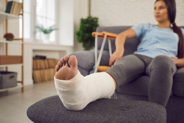
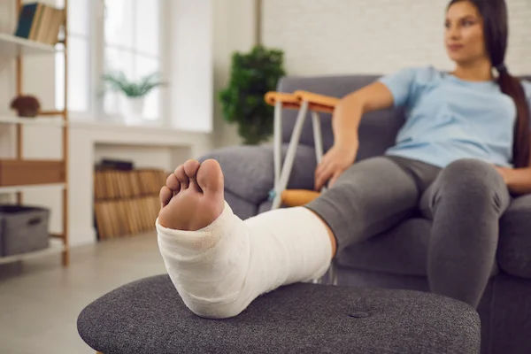
Hip fracture after a household fall
- A 76-year-old slips in the bathroom, can’t stand due to hip pain. X-rays show a displaced femoral neck fracture; surgery
is performed within 48 hours to improve outcomes. Post-op, she begins early mobilization and starts osteoporosis
evaluation/treatment. Lesson: Fall-proofing the home and screening for bone health can prevent or reduce severity.
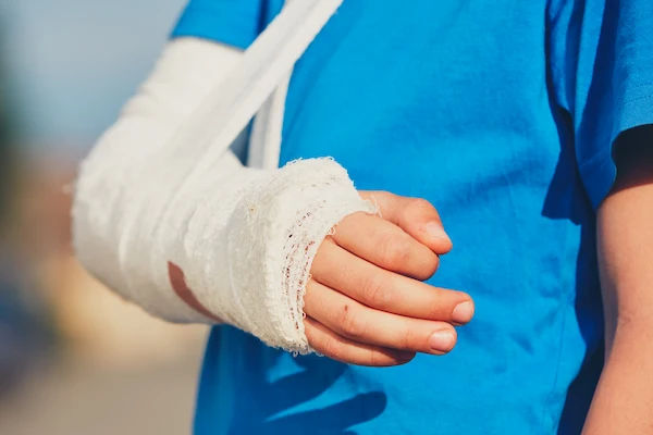
Child’s greenstick fracture on the playground
- A 7-year-old falls on an outstretched hand; wrist pain and swelling follow. X-ray shows a greenstick fracture of the
radius. A short period in a cast leads to excellent healing. Lesson: Children’s bones heal quickly, but growth plates must
be monitored.
Conclusion
Bone fractures range from tiny stress cracks to complex injuries that require surgery, but they share a common rule: prompt, appropriate care leads to better outcomes. Recognizing the signs of a fracture—sharp, localized bone pain, swelling, deformity, and trouble using the limb—helps you act quickly. Diagnosis typically starts with an X-ray, and treatment spans from splints and casts to advanced surgical fixation when alignment and stability demand it. The good news is that bones are built to heal. With the right plan, most fractures achieve solid union over weeks to months. Your everyday choices matter too—adequate protein, calcium, and vitamin D, plus smart, progressive activity and balance work—can shorten downtime and cut your risk of future injury. If symptoms persist or you’re worried about bone health, don’t wait. Talk to a clinician—Apollo24|7 can help with online consults, in-person appointments, and home collection for labs like vitamin D. A few practical steps now, like fall-proofing your home and building a strength routine, can safeguard your mobility and independence for years to come.
Consult a Top General Physician
Consult a Top General Physician

Dr. Ajay K Sinha
General Physician/ Internal Medicine Specialist
30 Years • MD, Internal Medicine
Delhi
Apollo Hospitals Indraprastha, Delhi
(225+ Patients)

Dr. Mijanur Rahaman Mondal
General Practitioner
3 Years • MBBS
Kolkata
Dr Utsa Basu Clinic, Kolkata
(25+ Patients)

Dr. Abhishek Gowda
General Physician/ Internal Medicine Specialist
3 Years • MBBS MD General Medicine
Bengaluru
PRESTIGE SHANTHINIKETAN - SOCIETY CLINIC, Bengaluru

Dr. Soumen Paul
General Practitioner
24 Years • MBBS
Kolkata
MCR SUPER SPECIALITY POLY CLINIC & PATHOLOGY, Kolkata
(25+ Patients)

Dr. Shashidhara Mahabala
General Physician/ Internal Medicine Specialist
28 Years • MBBS, MD, PGD (Diabetology)
Bengaluru
Apollo Clinic, HSR Layout, Bengaluru
(75+ Patients)
Consult a Top General Physician
Dr. Ajay K Sinha
General Physician/ Internal Medicine Specialist
30 Years • MD, Internal Medicine
Delhi
Apollo Hospitals Indraprastha, Delhi
(225+ Patients)
Dr. Mijanur Rahaman Mondal
General Practitioner
3 Years • MBBS
Kolkata
Dr Utsa Basu Clinic, Kolkata
(25+ Patients)
Dr. Abhishek Gowda
General Physician/ Internal Medicine Specialist
3 Years • MBBS MD General Medicine
Bengaluru
PRESTIGE SHANTHINIKETAN - SOCIETY CLINIC, Bengaluru
Dr. Soumen Paul
General Practitioner
24 Years • MBBS
Kolkata
MCR SUPER SPECIALITY POLY CLINIC & PATHOLOGY, Kolkata
(25+ Patients)
Dr. Shashidhara Mahabala
General Physician/ Internal Medicine Specialist
28 Years • MBBS, MD, PGD (Diabetology)
Bengaluru
Apollo Clinic, HSR Layout, Bengaluru
(75+ Patients)
More articles from Bone Fracture
.webp)
Frequently Asked Questions
Q1: How can I tell if I have a fracture or just a sprain?
Fracture pain is often sharp and localized over the bone, with swelling and sometimes deformity or inability to bear weight. Sprains affect ligaments around a joint and may allow limited use. Only an X-ray or MRI confirms it—if in doubt, seek care. If symptoms persist beyond two weeks, consult a doctor online with Apollo24|7.
Q2: What are the most common types of bone fractures?
Common types include transverse, oblique, spiral, and comminuted fractures. In kids, greenstick and buckle fractures are frequent. Overuse can cause stress fractures. Open fractures involve broken skin and require urgent care.
Q3: How long does a bone fracture take to heal?
Most uncomplicated fractures take 6–12 weeks to form solid bone, with continued remodeling for months. Children often heal faster; complex fractures may take longer. Good nutrition, not smoking, and following activity guidance help.
Q4: What are stress fracture symptoms?
Localized pain that worsens with activity and improves with rest, minimal swelling, and tenderness over a specific bone. Early X-rays can be normal; MRI is more sensitive. Activity modification is key to recovery.
Q5: How can I prevent osteoporosis-related fractures?
Combine calcium and vitamin D intake, weight-bearing and resistance exercises, fall prevention, and bone density screening (DEXA) when appropriate. Your doctor may recommend medication to strengthen bone. Apollo24|7 offers home collection for vitamin D testing.