Breast Calcifications: Types, Causes and Treatment
Understating what breast calcification is and Learning about its types, causes, its clinical inferences, diagnosis and treatment.

Written by Dr. Sonia Bhatt
Last updated on 3rd Jul, 2025
Introduction
Calcifications in breast tissue are tiny calcium deposits that show up as bright white spots on mammograms. They are quite common, especially in women after menopause, and are usually harmless. Since they are too small to be detected by touch, most individuals only find out about them during routine mammography.
Although the majority of calcifications are non-cancerous, certain features—like their size, shape, and pattern—can suggest whether further investigation is necessary. If the calcifications appear concerning, doctors may recommend additional imaging or a biopsy to rule out cancer. Regular screenings and discussions with a healthcare professional are important for maintaining breast health.
Types of Breast Calcifications
Breast calcifications are classified into two main types, each with unique appearances on a mammogram:
1.Macrocalcifications
These are large white spots that can be seen scattered throughout the breast tissue. Macrocalcifications are the most common form and are generally considered harmless, typically not requiring any follow-up imaging. Known as benign coarse calcifications, they can develop naturally as people age and are not associated with cancer. While macrocalcifications can occur at any age, they are more common after menopause. Possible causes include:
Calcium deposits in cysts or milk ducts due to aging
Previous injuries to the breast
Inflammation
2.Microcalcifications
These are small calcium deposits that present as tiny white dots on a mammogram. They often appear in areas of the breast where cell turnover is more rapid than usual. Although most microcalcifications are benign, a cluster of them might suggest:
Pre-cancerous changes
Early breast cancer
Causes of Breast Calcifications
Breast calcifications are common, particularly in individuals over 50, with about half of those assigned female at birth experiencing benign forms. The exact cause of these calcifications is not entirely clear, but factors contributing to their formation include:
Breast injuries
Breast cysts
Infections in the breast
Changes in blood vessels due to aging
Benign lumps (like fibroadenomas)
Mammary duct ectasia
Prior surgeries or treatments for breast cancer
While cancerous calcifications are often linked to ductal carcinoma in situ (DCIS), it’s crucial to understand that they are not caused by dietary calcium and do not evolve into breast cancer. Most calcifications are benign, though they can sometimes indicate underlying processes in breast tissue. Other benign causes may include past injuries, non-cancerous growths, previous radiation therapy, and calcium accumulation in blood vessels.
How Breast Calcifications are detected?
Most breast calcifications are benign and do not typically require further follow-up. However, there can be similarities between benign calcifications and those that may indicate an abnormality, as they can appear alike on a mammogram. For example, if a mammogram reveals a tight cluster of calcifications or small specks arranged in a line, the radiologist might suggest additional tests to rule out cancer.
Follow-up tests may include:
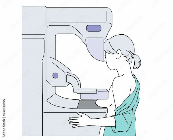
1. Diagnostic Mammogram: This type of mammogram provides more detailed images than a standard screening. It involves capturing images of the specific area of concern from various angles, enabling the radiologist to examine the calcifications more thoroughly. Magnified views are often employed to better evaluate the shape and arrangement of the calcifications.
2. Biopsy: In certain instances, a biopsy may be needed to ascertain the cause of the calcium deposits. This minimally invasive procedure utilises imaging to guide the removal of a small sample of breast tissue for analysis by a pathologist. The biopsy is generally performed as a stereotactic procedure, where mammogram images assist in extracting small tissue samples from the area of concern, often using a core needle.
These follow-up tests are crucial in determining whether the calcifications are benign or if further investigation is warranted.
Interpretation of Breast Calcifications
Evaluating breast calcifications involves examining their shape, distribution, and any changes over time. The shape is crucial for determining if they are benign or potentially malignant. If calcifications are not clearly benign or high-risk, they are classified as of intermediate concern, often necessitating a biopsy to identify the underlying cause.
1. Morphology: The shape of the calcifications is essential for differentiating between benign and malignant conditions. If they cannot be easily classified, they are termed of intermediate concern. Descriptions should include shape and distribution, following BI-RADS guidelines.
2. Distribution: The BI-RADS atlas provides descriptions for calcification distribution:
3. Diffuse or Scattered: Widely distributed calcifications or multiple similar clusters, typically benign.
4. Regional: Calcifications spread across a larger volume of tissue (over 2 cc), usually indicating benignity.
5. Clustered: At least five calcifications in a small area; linear arrangements may suggest ductal deposits.
6. Segmental: Calcifications located in ducts and branches of a specific segment, potentially indicating malignancy.
Diffuse or scattered distributions are generally benign, while segmental patterns may suggest malignancy. Distinguishing between regional and segmental can be difficult due to unclear boundaries on imaging.
Clustered calcifications can indicate either benign or malignant conditions. Scattered clusters are more likely benign, whereas a single cluster raises suspicion for malignancy. Linear distributions are often associated with ductal carcinoma in situ (DCIS), where calcifications fill the entire duct and its branches.
Risk Factors Associated with Breast Calcifications
If you have previously been diagnosed with breast cancer or have a higher risk due to a strong family history or genetic mutation, you may be particularly concerned about calcifications found on your mammogram. Treatments for cancer, such as surgery, reconstruction, and radiation therapy, can lead to tissue damage and scarring, which might cause calcifications to appear on mammograms.
Diagnostic Procedures for Unusual Calcifications
If calcifications are assessed as benign, further testing is usually unnecessary. However, if they are deemed indeterminate or suspicious, additional investigations will be required for an accurate diagnosis.
1. Mammogram: You may have a follow-up mammogram known as tomosynthesis, which provides a more detailed image of the affected area.
2. Ultrasound Scan: An ultrasound scan uses sound waves to produce images of the breast tissue.
3. Breast Biopsy: A biopsy may be needed to collect tissue samples for examination. Common types include:
4. Core Biopsy: A hollow needle is used to extract a small sample of breast tissue, guided by a mammogram or ultrasound. This is done under local anaesthetic, and the samples are sent for analysis.
5. Vacuum-Assisted Biopsy: Similar to a core biopsy but allows for multiple samples to be collected through a single incision using a vacuum device. This also uses local anaesthetic.
6. Vacuum-Assisted Excision Biopsy: This procedure removes a larger area of calcification and may help you avoid general anaesthetic.
7. Inserting a Metal Marker: Sometimes, a small metal clip is placed in the breast at the biopsy site to aid in locating the area for future procedures. The titanium clip is safe to leave in and won’t trigger airport security alarms and most clips are MRI-compatible.
Treatment Options for Concerning Calcifications
In most instances, no treatment is required for breast calcifications.
1. Benign Calcifications: These are harmless and do not need further evaluation.
2. Probably Benign Calcifications: With less than a 2% chance of being cancerous, these are usually monitored every six months for at least a year. If no changes are detected during this time, your doctor may suggest annual mammograms.
3. Suspicious Calcifications: These may indicate the presence of early cancer, leading your doctor to recommend a biopsy to collect a small tissue sample for examination. If cancer is confirmed, treatment may involve surgery to remove the affected tissue.
Types of Biopsies
1. Core Needle Biopsy: This is performed under local anaesthetic, using a thin, hollow needle guided by imaging to obtain a small sample of tissue.
2. Surgical Biopsy: If the core needle biopsy does not yield clear results, a surgical biopsy may be necessary. This procedure is done under local or general anaesthetic, with a radiologist marking the area for the surgeon to extract the sample for analysis.
When is Surgery required?
Surgery may be necessary if:
A biopsy is not feasible.
A biopsy does not provide clear results.
Findings suggest atypia (unusual changes).
Results indicate early cancer.
Surgery is typically performed under general anaesthetic, allowing for same-day discharge.
Preventive Measures and Health Recommendations
If your radiologist suspects that your breast calcifications may be linked to precancerous changes or breast cancer, further assessments will be necessary. This may include another mammogram with magnified views for a more detailed examination, or a biopsy to analyse a tissue sample.
The radiologist might also ask for previous mammogram images to compare and determine if the calcifications are new or have changed in number or appearance.
If the calcifications are believed to be due to a benign condition, a follow-up mammogram with magnified views may be recommended in six months. During this follow-up, they will look for any alterations in the shape, size, and quantity of calcifications, or confirm that they have remained stable.
Conclusion
Breast calcifications are common and often result from noncancerous factors like previous surgery, infections, or natural ageing. If calcifications are found, your doctor will advise you on any necessary follow-up tests or treatments. It’s natural to feel anxious about the possibility that calcifications might indicate cancer. However, it’s important not to jump to conclusions if your provider finds them on a mammogram. Many benign conditions can cause calcium deposits in breast tissue, which usually do not suggest malignancy. If cancer is diagnosed, discussing your prognosis with your provider is essential, as effective treatments may be available.
Consult Top Oncologist
Consult Top Oncologist

Dr. Tarun Jindal
Uro Oncologist
14 Years • MS (AIIMS, New Delhi), MCh (Gold Medalist), Fellow, VUI, Henry Ford Hospital, Detroit, USA; Robotic and Laparoscopic surgeon
Kolkata
Apollo Multispeciality Hospitals , Kolkata, Kolkata
(100+ Patients)

Dr Nikhil Suresh Ghadyalpatil
Oncologist
18 Years • MBBS, MD (G. Med), DNB (G.Med), MNAMS DM (Medical Oncology - Tata Memorial Hospital) European Certification In Medical Oncology (ECMO) MRCP (Med Onco SCE), PDCR
Hyderabad
Apollo Hospitals Jubilee Hills, Hyderabad

Dr. Sandeep Muzumder
Radiation Specialist Oncologist
21 Years • MBBS (JIPMER, Pondicherry), MD (AIIMS, New Delhi)
Bhubaneswar
Apollo Hospitals Old Sainik School Road, Bhubaneswar

Dr Devashish Tripathi
Radiation Specialist Oncologist
20 Years • MBBS, PLAB, MRCP (UK)- General Medicine, FRCR (Oncology), Certificate of Completion of Training (CCT)- Clinical Oncology
Delhi
Apollo Hospitals Indraprastha, Delhi

Dr. Gopal Kumar
Head, Neck and Thyroid Cancer Surgeon
15 Years • MBBS, MS , FARHNS ( Seoul, South Korea ), FGOLF ( MSKCC, New York )
Delhi
Apollo Hospitals Indraprastha, Delhi
(25+ Patients)