
Chondrosarcoma Cancer: Signs and Symptoms
Learn about chondrosarcoma, a rare type of bone cancer, including its signs, symptoms, and when to seek medical attention for early detection.

Written by Dr. Md Yusuf Shareef
Reviewed by Dr. Dhankecha Mayank Dineshbhai MBBS
Last updated on 13th Jan, 2026
Introduction
Chondrosarcoma is a rare type of cancer that originates in the cartilage cells. While the word "cancer" can be alarming, understanding this specific disease is the first step toward empowerment. Unlike many cancers, chondrosarcoma most commonly appears in adulthood and has a spectrum of behaviors, from slow-growing and manageable to more aggressive forms. The challenge often lies in its early detection, as its initial signs can be subtle and mistaken for common aches and pains. This article will guide you through the key signs and symptoms of chondrosarcoma, helping you distinguish between everyday discomfort and something that warrants a deeper look with a healthcare professional. We'll explore where it occurs, what it feels like, and the crucial steps toward diagnosis and treatment.
What exactly is Chondrosarcoma?
Chondrosarcoma is a type of sarcoma, a broad category of cancers that develop in the bones and soft tissues. Specifically, it arises from transformed cartilage cells. Cartilage is the tough, flexible connective tissue that cushions your joints and plays a key role in bone development. When these cells mutate and grow uncontrollably, they form a malignant tumour known as chondrosarcoma.
How Chondrosarcoma Differs from Other Bone Cancers
It's important to distinguish chondrosarcoma from more common bone cancers like osteosarcoma (which starts in bone cells) or Ewing sarcoma (which often affects children and adolescents). Chondrosarcoma typically occurs in older adults (over 40) and is known for its production of cartilaginous matrix, which is a hallmark feature visible on imaging and under a microscope.
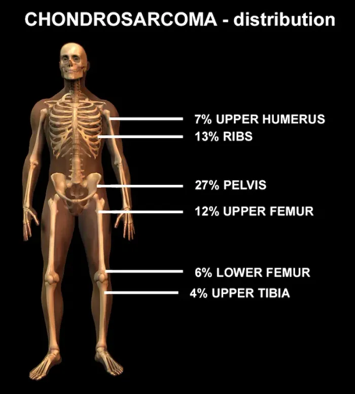
Common Locations in the Body
This cancer has a preference for certain areas of the skeleton. The most common sites include:
• The pelvis and hip bones
• The shoulder (proximal femur and humerus)
• The ribs
• The spine and skull base
Understanding these locations helps contextualise the symptoms, as pain in the hip or shoulder could be more than just arthritis.
Consult an Orthopaedic Oncologist for the best advice
The Early Warning Signs: Listening to Your Body
Early detection significantly improves outcomes. The symptoms often develop slowly and insidiously, which is why they are frequently dismissed.
The Primary Symptom: A Persistent Pain
The most common and often the first symptom of chondrosarcoma is a deep, dull ache. Unlike the pain from a minor injury that fades, this pain:
• Is persistent and progressive, often worsening over weeks or months.
• May be more noticeable at night or during rest, which is unusual for typical muscle strain.
• Is localised to one specific area, like the pelvis or a long bone in the arm or leg.
The Telling Sign: A Noticeable Lump or Mass
As the tumour grows, it may form a palpable lump or swelling. This mass:
• Is firm and attached to the underlying bone.
• May be tender or painful to the touch.
• Can interfere with the normal function of a nearby joint, making it difficult to move your arm or walk normally if it's near a hip or shoulder.
Swelling and Tenderness Around a Joint
Localised swelling and tenderness around a joint without a history of injury is a significant red flag. This is often seen in cases of chondrosarcoma in the pelvis or shoulder, where the tumour causes inflammation in the surrounding tissues.
Advanced Symptoms and Potential Complications
If left undiagnosed, the tumour can grow and lead to more severe symptoms.
Neurological Symptoms (Spinal or Pelvic tumours)
Tumours located in the spine or pelvis can press on nerves or the spinal cord. This can cause:
• Sciatica-like pain radiating down a leg.
• Numbness, tingling, or weakness in the limbs.
• In severe cases, loss of bowel or bladder control (a medical emergency).
Pathological Fractures: When Bones Break Unexpectedly
A growing tumour weakens the bone from within. A pathological fracture occurs when the bone breaks during normal, everyday activity, like a minor stumble or even just bearing weight, because it has been structurally compromised by the cancer. This is a serious complication that requires immediate medical attention.
What Causes Chondrosarcoma? Understanding Risk Factors
The exact cause of most chondrosarcomas is unknown. They often appear spontaneously. However, certain risk factors and pre-existing conditions can increase susceptibility.
Pre-existing Benign Conditions
In some cases, a benign cartilage tumour can undergo malignant transformation. The most common example is an enchondroma (a non-cancerous cartilage tumour inside the bone) or an osteochondroma (a bony outgrowth capped with cartilage). Patients with multiple benign tumours, a condition called Ollier's disease or Maffucci syndrome, have a significantly higher lifetime risk of developing chondrosarcoma.
Genetic Syndromes and Family History
While most cases are not inherited, certain rare genetic syndromes are linked to a higher incidence of sarcomas. However, for the vast majority of patients, there is no clear family history or genetic cause.
How is Chondrosarcoma Diagnosed? The Medical Journey
If chondrosarcoma is suspected based on symptoms, a doctor will initiate a diagnostic workup.
The Crucial First Step: Imaging Scans (X-ray, MRI, CT)
• X-ray: Often the first test ordered. It can reveal bone destruction, calcification patterns typical of cartilage tumours, and soft tissue masses.
• MRI (Magnetic Resonance Imaging): This is the most valuable tool for defining the tumour. It shows the exact size, location, and extent of the tumour within the bone and soft tissues and its relationship to nerves and blood vessels.
• CT Scan: Useful for evaluating the chest for potential spread (metastasis) and for assessing the bone detail in complex areas like the pelvis.
The Gold Standard: Biopsy and Histological Grading
A biopsy, where a small sample of the tumour is removed and analysed by a pathologist, is the only way to confirm a chondrosarcoma diagnosis. The biopsy also determines the tumour's grade (low, intermediate, or high), which describes how aggressive the cancer cells appear and is the most critical factor in determining treatment and prognosis.
Treatment Pathways: From Surgery to Monitoring
The cornerstone of treatment is surgery. The goal is to remove the entire tumour while preserving as much function as possible.
Wide Local Excision: The Primary Curative Treatment
This surgical procedure involves removing the tumour along with a surrounding margin of healthy tissue to ensure no cancer cells are left behind. For limb tumours, limb salvage surgery is almost always possible, where the tumour is removed and the bone is reconstructed using a metal prosthesis or a bone graft.
Addressing Advanced or Aggressive Cases
Radiation Therapy: May be used if the tumour cannot be fully removed with clean margins (e.g., in the base of the skull) or to treat recurrent disease.
Chemotherapy: Traditional chemotherapy is generally not effective for most chondrosarcomas. However, it may be considered for the very rare, highly aggressive dedifferentiated subtype.
If you are experiencing persistent bone pain or a growing lump, it is crucial to consult a specialist.
Prognosis and Long-Term Outlook
The prognosis for chondrosarcoma is generally favorable, especially for low-grade tumours that are completely surgically removed. The chondrosarcoma survival rate is highly dependent on the grade, location, and size of the tumour at diagnosis, and whether it has spread. High-grade tumours have a higher risk of recurrence and metastasis, most commonly to the lungs. Long-term, regular follow-up with imaging is essential to monitor for any signs of recurrence.
Conclusion
Recognising the signs of chondrosarcoma cancer, primarily a deep, persistent pain and a growing mass, can lead to earlier diagnosis and more successful treatment outcomes. While the journey from symptom to diagnosis can be daunting, modern medical advances, particularly in imaging and surgical techniques, offer effective management for this disease. It's vital to listen to your body and not dismiss ongoing pain as mere aging or a simple injury. If your symptoms are persistent and unexplained, proactively seeking a medical opinion is the most important step you can take. Early intervention is key to preserving your health, mobility, and quality of life.
Consult an Orthopaedic Oncologist for the best advice
Consult an Orthopaedic Oncologist for the best advice

Dr Gowshikk Rajkumar
Oncologist
10 Years • MBBS, DMRT, DNB in Radiation oncology
Bengaluru
Apollo Clinic, JP nagar, Bengaluru

Dr Devashish Tripathi
Radiation Specialist Oncologist
20 Years • MBBS, PLAB, MRCP (UK)- General Medicine, FRCR (Oncology), Certificate of Completion of Training (CCT)- Clinical Oncology
Delhi
Apollo Hospitals Indraprastha, Delhi
(25+ Patients)
Dr. B Shravanthi Reddy
Radiation Specialist Oncologist
8 Years • MBBS, DNB(Radiation Oncology)
Manikonda Jagir
Apollo Clinic, Manikonda, Manikonda Jagir

Dr. Subhash Chandra Chanana
Oncologist
51 Years • M.B.B.S , M.S. (General Surgery), F.A.C.S (Oncosurgeon)
Gurugram
APOLLO SUGAR CLINICS GURUGRAM, Gurugram

Dr. V R N Vijay Kumar
Surgical Oncologist
9 Years • MBBS, MS (Gen. Surg.), DNB (Surg. Onco.)
Ahmedabad
Apollo Hospitals Gandhinagar, Ahmedabad
Consult an Orthopaedic Oncologist for the best advice

Dr. Preetham Raj Chandran
Orthopaedician
10 Years • MBBS, MS (Orthopedics), FIASM, FIJR
Bangalore
Apollo Clinic Bellandur, Bangalore

Dr. Sushruth J
Orthopaedician
5 Years • MBBS, MS (ORTHOPEDICS),Fellowship in Arthroplasty,FRGUHS – Spine surgery,FIFA Diploma in Football Medicine
Bengaluru
Apollo Clinic, JP nagar, Bengaluru

Dr Abhishek Nandi
Paediatric Orthopaedician
10 Years • MBBS, M.S. ortho (JIPMER), Fellow Paed Ortho (CMC Vellore), Post- Doctoral Fellow Paed Ortho( MGR U) Speciality- Paediatric fractures, Bone and joint infection, Deformity correction, Bony tumour, Cerebral Palsy, Limb Reconstruction
Kolkata
Apollo Multispeciality Hospitals , Kolkata, Kolkata
Dr. B Shravanthi Reddy
Radiation Specialist Oncologist
8 Years • MBBS, DNB(Radiation Oncology)
Manikonda Jagir
Apollo Clinic, Manikonda, Manikonda Jagir

Dr Navneet Saraiya
Spine Surgeon
19 Years • MBBS, MRCS Edinburgh, FRCS Trauma & Orthopaedics, FRCS Glasgow
Ahmedabad
Apollo Hospitals Gandhinagar, Ahmedabad
(25+ Patients)
Consult an Orthopaedic Oncologist for the best advice
Dr. Preetham Raj Chandran
Orthopaedician
10 Years • MBBS, MS (Orthopedics), FIASM, FIJR
Bangalore
Apollo Clinic Bellandur, Bangalore
Dr. Sushruth J
Orthopaedician
5 Years • MBBS, MS (ORTHOPEDICS),Fellowship in Arthroplasty,FRGUHS – Spine surgery,FIFA Diploma in Football Medicine
Bengaluru
Apollo Clinic, JP nagar, Bengaluru
Dr Abhishek Nandi
Paediatric Orthopaedician
10 Years • MBBS, M.S. ortho (JIPMER), Fellow Paed Ortho (CMC Vellore), Post- Doctoral Fellow Paed Ortho( MGR U) Speciality- Paediatric fractures, Bone and joint infection, Deformity correction, Bony tumour, Cerebral Palsy, Limb Reconstruction
Kolkata
Apollo Multispeciality Hospitals , Kolkata, Kolkata
Dr. B Shravanthi Reddy
Radiation Specialist Oncologist
8 Years • MBBS, DNB(Radiation Oncology)
Manikonda Jagir
Apollo Clinic, Manikonda, Manikonda Jagir
Dr Navneet Saraiya
Spine Surgeon
19 Years • MBBS, MRCS Edinburgh, FRCS Trauma & Orthopaedics, FRCS Glasgow
Ahmedabad
Apollo Hospitals Gandhinagar, Ahmedabad
(25+ Patients)
Frequently Asked Questions
1. Is chondrosarcoma curable?
Yes, especially low-grade chondrosarcoma is often curable with complete surgical removal. The cure rate is highest when the tumour is detected early and before it has spread.
2. What does chondrosarcoma pain feel like?
The pain is typically a deep, dull, and persistent ache within the bone. It is often described as worsening at night or during rest, which distinguishes it from pain caused by physical activity or arthritis.
3. Can a benign cartilage tumour turn into chondrosarcoma?
Yes, in rare cases, pre-existing benign tumours like enchondromas or osteochondromas can undergo malignant transformation into chondrosarcoma, particularly in individuals with associated genetic syndromes.
4. How fast does chondrosarcoma grow?
The growth rate varies significantly by grade. Low-grade chondrosarcomas are very slow-growing, sometimes taking years to become symptomatic. High-grade tumours can grow much more rapidly.
5. Does chondrosarcoma show up on an X-ray?
Yes, an X-ray is often the first test to suggest a bone tumour. It can show characteristic patterns of bone destruction and calcification that raise suspicion for a cartilage-based tumour like chondrosarcoma, though an MRI is needed for full evaluation.