

White Spots On Skin Reasons, Types, And Fixes
Discover the common reasons for white spots on skin, their types, and effective ways to manage or treat them. Learn when to seek medical advice for skin changes.

Written by Dr. Mohammed Kamran
Reviewed by Dr. D Bhanu Prakash MBBS, AFIH, Advanced certificate in critical care medicine, Fellowship in critical care medicine
Last updated on 13th Jan, 2026
Introduction
Noticing a white spot on your skin can be worrying—especially if it spreads or stands out in photos or bright light. The good news: most white spots on skin have clear reasons and effective treatments once you know what you’re dealing with. In this practical guide, we explain the most common reasons for white spots on skin, how to tell them apart at home, and what steps you can take right away. We’ll cover everyday causes like tinea (pityriasis) versicolor and pityriasis alba, as well as vitiligo, idiopathic guttate hypomelanosis, and post-inflammatory hypopigmentation. You’ll learn how to use simple clues—like scale, borders, and location—to narrow the possibilities, plus what tests a clinician might perform (such as a Wood’s lamp or KOH skin scraping). We’ll also talk through treatments that work, prevention tips, skin-of-colour considerations, and when to seek medical care. If symptoms persist beyond two weeks or you’re unsure about the reason for white spots on skin, consult a doctor online with Apollo 24|7 for further evaluation and guidance.
What Counts as a “White Spot” on Skin?
White spots aren’t all the same. Some are truly depigmented (loss of all pigment), while others are hypopigmented (reduced pigment) or appear lighter for other reasons (like scaling that reflects light). This distinction matters because treatments and expectations differ.
Consult Top Specialists
Hypopigmented vs. depigmented vs. hypochromic
Hypopigmented lesions have less melanin but not zero; they often look lighter rather than stark white. Depigmented lesions (as in vitiligo) can be “paper white,” with sharp borders and sometimes hair turning white in the area. Hypochromic often refers to subtler lightness. Under a Wood’s lamp, depigmented patches tend to show bright fluorescence, aiding diagnosis.
Texture clues
Run a fingertip across the area. If it’s slightly scaly or flaky, a superficial fungal overgrowth like tinea versicolor is more likely. Smooth patches with sharp borders raise suspicion for vitiligo; dry, slightly rough patches on kids’ cheeks often suggest pityriasis alba.
Location and number
Face patches in children often mean pityriasis alba. Scattered tiny spots on shins and forearms in adults can point to idiopathic guttate hypomelanosis (IGH). Multiple light macules on the upper back and chest in hot weather may be tinea versicolor. Genital or perianal white plaques could be lichen sclerosus and need medical assessment.
The Fast Filter: A Simple Flow to Narrow the Reason
Here’s a quick way to sort common causes at home before you see a clinician.
Step 1: Scale or no scale?
If gently scraping the area with a fingernail reveals fine powdery scale, tinea versicolor or pityriasis alba climbs the list. Tinea versicolor often has subtle scale; pityriasis alba tends to be dry and matte. No scale? Consider vitiligo, IGH, post-inflammatory hypopigmentation.
Step 2: Borders and shape
Sharp, map-like borders with stark white colour favour vitiligo. Fuzzy edges that blend into surrounding skin suggest post-inflammatory hypopigmentation or pityriasis alba. Tiny, round “confetti” spots on sun-exposed limbs suggest IGH.
Step 3: Distribution and timing
Trunk/shoulders after heat/sweat: tinea versicolor. Cheeks in kids with eczema: pityriasis alba. Hands, around eyes/mouth, or body folds with symmetry: vitiligo. After a healed rash or burn: post-inflammatory hypopigmentation.
If you can’t confidently narrow it or if lesions are on genitals, rapidly spreading, or symptomatic (itch/pain), book a clinician visit. If symptoms persist beyond two weeks, consult a doctor online with Apollo 24|7 for further evaluation.
Tinea Versicolor (Pityriasis Versicolor): The Common Fungal Cause
Tinea versicolor is caused by an overgrowth of Malassezia yeast that normally lives on skin. In warm, humid climates or after sweating, it can shift into a form that interferes with pigment production, creating lighter (or sometimes darker) patches with fine scale. It commonly affects the trunk, shoulders, neck, and upper arms.
Signs and seasonality
Look for multiple, slightly scaly patches that may coalesce into larger areas. Tan-to-pale patches that look more obvious after sun exposure are typical. Many people notice a summer pattern: more visible in hot months and improved in cooler weather.
Diagnosis
Clinicians can confirm tinea versicolor with:
• KOH (potassium hydroxide) skin scraping that shows characteristic “spaghetti and meatballs” yeast and hyphae.
• Wood’s lamp sometimes shows a yellow-green fluorescence, but this is variable and not required.
At home, the presence of fine scale and typical distribution, plus response to antifungal shampoo, are clues. If unsure, avoid steroid creams; they won’t treat the yeast.
Treatment and relapse prevention
First-line options include:
• Topical antifungal shampoos (selenium sulfide 2.5%, ketoconazole 2%) used as body washes, left on 5–10 minutes before rinsing, daily for 5–7 days, then weekly for prevention.
• Topical antifungal creams (clotrimazole, ketoconazole) for limited areas.
• Oral antifungals (itraconazole/fluconazole) may be prescribed for extensive or recurrent cases—see a doctor for this.
Note: Skin colour can take weeks to months to normalise after yeast is controlled; that’s expected. For frequent relapses, monthly maintenance washes help. Related term to include: KOH test for tinea versicolour.
Pityriasis Alba: The Post-Eczema Fade on Kids’ Cheeks
Pityriasis alba is common in children and teens, particularly those with eczema or sensitive skin. It presents as light patches—usually on the face (cheeks), sometimes upper arms—with faint scale and ill-defined borders. It’s more obvious after sun exposure because surrounding skin tans more than the patches.
Differentiating from vitiligo
Pityriasis alba patches are not paper white; borders are soft and the patches are matte/dry. Vitiligo is whiter, with sharper edges, and may involve areas around the eyes and mouth. A Wood’s lamp does not show the striking fluorescence seen in vitiligo.
Care and treatments
Gentle skincare is the cornerstone:
• Daily bland moisturisers to repair the barrier.
• Mild hydrocortisone or calcineurin inhibitors (pimecrolimus/tacrolimus) for short courses if eczema is active, per clinician advice.
• Sunscreen to reduce contrast and make patches less visible.
Patches often resolve over months. If not improving, or if sharply defined white patches appear, see a dermatologist. Related term to include: white spots on children’s face.
Vitiligo: Autoimmune White Patches with Sharp Borders
Vitiligo results from immune-mediated loss of melanocytes (pigment cells), leading to chalk-white patches with well-defined edges. It can begin at any age and occurs worldwide, affecting about 0.5–2% of people.
Early signs and spread patterns
Common areas include hands, around body openings (eyes, mouth), elbows, knees, and genital regions. Hair in the patch may turn white (poliosis). Patches can be symmetrical (generalised vitiligo) or segmental (limited to one side). Sunburn can worsen the contrast, so sun protection is key.
Workup and why thyroid screening matters
Vitiligo is associated with other autoimmune conditions, particularly autoimmune thyroid disease. Clinicians may consider thyroid function tests and antibodies as part of the evaluation. A Wood’s lamp exam helps distinguish depigmented patches from hypopigmented ones. If labs are needed, Apollo 24|7 offers convenient home collection for tests like thyroid profiles and vitamin D (if clinically indicated).
Consult Top Specialists
Treatment options
• Topical corticosteroids or calcineurin inhibitors for limited patches.
• Phototherapy (narrowband UVB, excimer) for more extensive disease.
• Emerging therapies (e.g., topical ruxolitinib in some regions) show promise.
• Camouflage cosmetics and skin dyes can reduce visibility.
Vitiligo is not contagious. Early treatment improves the chance of repigmentation. If you notice new, rapidly spreading white patches, consult a doctor online with Apollo 24|7 for guidance and possible referral.
Idiopathic Guttate Hypomelanosis: Tiny “Confetti” Spots on Sun-Exposed Areas
IGH presents as multiple small, round or oval, 2–5 mm white macules on sun-exposed areas—forearms, shins, and sometimes shoulders. It becomes more common with age and cumulative sun exposure.
Why prevention matters
IGH is benign but persistent. UV exposure likely plays a role, so daily broad-spectrum sunscreen and protective clothing help reduce new lesions. Moisturisers won’t reverse IGH, but may improve overall skin tone.
Cosmetic options and expectations
Treatments aim to reduce contrast, not “cure”:
• Targeted procedures (e.g., fractional lasers, excimer) may help some patients; results vary, and multiple sessions may be needed.
• Self-tanning lotions or cosmetic camouflage can blend tone effectively in the short term.
Discuss realistic outcomes with a dermatologist.
Post-Inflammatory Hypopigmentation: Light Marks After a Rash or Injury
After inflammatory skin conditions (eczema, psoriasis, acne) or minor injuries, healing can leave areas with reduced pigment. These are usually lighter with ill-defined borders and gradually repigment over weeks to months as melanocytes recover.
Common triggers
Eczema flares, psoriasis plaques, insect bites, dermatitis, friction, or chemical irritation may precede the light spots. On darker skin tones, this contrast can be pronounced and persist longer.
What you can do
• Treat the underlying condition to prevent new spots.
• Gentle skin care (fragrance-free cleansers, moisturisers).
• Sun protection to reduce contrast and prevent tanning of surrounding skin.
If areas don’t improve over several months or the diagnosis is unclear, a clinician may recommend a Wood’s lamp exam or, rarely, a biopsy to exclude other conditions. For persistent or unclear cases, consult a doctor online with Apollo 24|7; if needed, book a physical visit for examination and tests.
Less Common but Important Causes
Here are some more less common causes:
Lichen sclerosus
This inflammatory condition causes white, thin, sometimes shiny patches—most often in the genital or perianal region—and can cause itching, pain, or tearing. It requires medical treatment (often potent topical steroids) to reduce symptoms and prevent scarring.
Pityriasis lichenoides chronica and hypopigmented mycosis fungoides
Rare, chronic conditions that can present with hypopigmented patches, especially in children or adolescents with darker skin tones (HMF). These require a dermatologist evaluation; biopsy may be necessary.
Genetic and rare causes
Piebaldism (stable depigmented patches present from birth), tuberous sclerosis “ash leaf” spots (oval hypopigmented macules), and others usually have distinctive features and associated findings. Medical evaluation is essential for these.
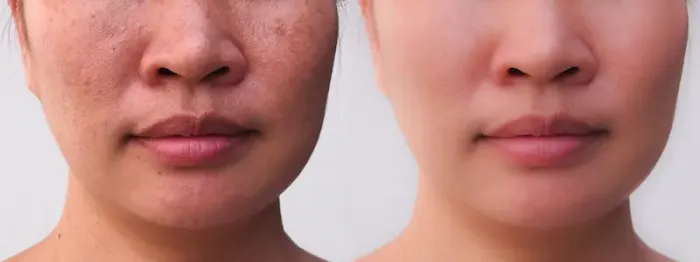
Skin Colour Considerations
White spots can look more prominent on darker skin tones due to higher background contrast. Post-inflammatory hypopigmentation and pityriasis alba may persist longer and be more visible.
• Reducing contrast
Daily broad-spectrum sunscreen (SPF 30+) prevents surrounding skin from tanning more than the spot. Tinted sunscreens can help blend tone.
• Pigment-protective routines
Barrier repair (ceramides), anti-inflammatory care for eczema-prone skin, and cautious use of any bleaching or exfoliating agents reduce the risk of new hypopigmented or hyperpigmented marks.
When to See a Doctor?
Seek care if:
• Rapidly spreading white patches
• Involvement of eyes, mouth, hands, genitals
• Itching, pain, fissuring, or bleeding
• Uncertain diagnosis or no improvement with simple measures in 2–4 weeks
Treatments That Work: Based on the Reason
Here are some treatment options:
• Antifungals for tinea versicolor
Ketoconazole or selenium sulfide washes; clotrimazole creams; occasional oral therapy for recurrences. Maintenance washes may prevent relapse.
• Anti-inflammatory topicals
Low-potency steroids or calcineurin inhibitors help pityriasis alba or post-inflammatory hypopigmentation by calming background eczema/dermatitis.
• Vitiligo therapies
Topical steroids/calcineurin inhibitors, narrowband UVB, excimer laser, and, in select regions, topical JAK inhibitors (e.g., ruxolitinib) can promote repigmentation. Cosmetic camouflage offers immediate blending.
• Camouflage and colour correction
Dermatologist-recommended camouflage cosmetics, self-tanners, and skin dyes can restore colour match for events or everyday life.
Everyday Care and Prevention Tips
Here’s how you can prevent it:
• Sun protection: SPF 30+ broad-spectrum sunscreen daily on exposed areas. Reapply during outdoor activities. Hats and UPF clothing add protection and reduce contrast.
• Gentle skin care: Fragrance-free cleansers, ceramide-rich moisturisers, lukewarm water, and brief showers. Avoid harsh scrubs and hot water that can worsen dryness and scaling.
• Tinea versicolor relapse prevention: Monthly antifungal wash (ketoconazole or selenium sulfide) during warm months and after workouts can reduce recurrences. Launder workout clothes promptly and shower after heavy sweating.
Myths vs Facts About White Spots
• Myth: “White spots mean calcium/vitamin deficiency.”
Fact: Most white spots are unrelated to calcium. Some clinicians may check vitamin D in certain contexts, but deficiencies are not the usual reason for white spots on skin.
• Myth: “They’re contagious.”
Fact: Vitiligo, pityriasis alba, IGH, and post-inflammatory hypopigmentation are not contagious. Tinea versicolor involves yeast that lives on everyone’s skin; overgrowth is not “caught” like ringworm but can flare with heat/sweat.
• Myth: “Sun will fix it.”
• Fact: Sun darkens surrounding skin more than the light spot, increasing contrast and sometimes triggering flares.
Emotional Well-Being and Support
Visible skin changes can affect self-esteem and social interactions. Consider:
• Camouflage techniques for events or photos.
• Support groups for vitiligo or chronic skin conditions.
• Counselling if distress impacts daily life.
Talk openly with your healthcare provider; cosmetic support is meaningful care, not vanity.
For Parents: White Spots on Children’s Skin
Common causes include pityriasis alba and post-inflammatory hypopigmentation after eczema or rashes. Gentle care and sun protection are key. See a paediatrician or dermatologist if:
• Patches are sharply white with well-defined borders (possible vitiligo).
• There’s itch, pain, or cracking.
• No improvement over a few months with moisturising and sunscreen.
If your child’s white patches persist or spread, consult a doctor online with Apollo 24|7 for guidance and, if needed, a referral.
Consult Top Specialists
Conclusion
White spots on skin can be surprising, but most have straightforward reasons and proven solutions. By looking for simple clues—scale, borders, location, and timing—you can often narrow the possibilities. Tinea versicolor commonly causes scaly light patches on the trunk that respond well to antifungal washes. Pityriasis alba often affects children’s faces and improves with moisturisers and sunscreen. Vitiligo presents as sharply bordered white patches and benefits from early, targeted treatment and sun protection. Tiny “confetti” spots on sun-exposed limbs often reflect idiopathic guttate hypomelanosis, where prevention and cosmetic blending are the mainstays. Post-inflammatory hypopigmentation is common after rashes or injuries and typically repigments over time with supportive care.
When in doubt—or if lesions are spreading quickly, symptomatic, or in sensitive areas—professional evaluation is the safest next step. A clinician may use simple tools like a Wood’s lamp or KOH scraping to confirm the reason and guide treatment. If you need guidance, consult a doctor online with Apollo 24|7. If lab tests are recommended (for example, thyroid tests in suspected vitiligo), Apollo 24|7 provides convenient home collection. With the right information, a practical plan, and consistent skin care, you can reduce contrast, support repigmentation, and feel more confident in your skin.
Consult Top Specialists
Dr. Mayuri Jain
Dermatologist
11 Years • MBBS, MD Dermatology , Venereology & Leprosy
Delhi
Dr Mayuri Jain Clinic, Delhi
Dr. Kavitha Killaparthy
Dermatologist
23 Years • MBBS,DIPLOMA(DERMATOLOGY,VENEREOLOGY,LEPROSY)
Hyderabad
JDS Skin & Hair Clinic, Hyderabad

Dr Ekansh Shekhar
Dermatologist
10 Years • MBBS MD
Lucknow
Apollo Clinic Hazratganj, Lucknow

Dr. Ashwini T
Dermatologist
8 Years • MBBS, MD ( DERMATOLOGY )
Bengaluru
Apollo Clinic, JP nagar, Bengaluru

Dr. Soumini S
Dermatologist
9 Years • MBBS, MD DERMATOLOGY,VENEREOLOGY, LEPROLOGY
Bengaluru
Apollo Medical Center, Marathahalli, Bengaluru
Consult Top Specialists
Dr. Mayuri Jain
Dermatologist
11 Years • MBBS, MD Dermatology , Venereology & Leprosy
Delhi
Dr Mayuri Jain Clinic, Delhi
Dr. Kavitha Killaparthy
Dermatologist
23 Years • MBBS,DIPLOMA(DERMATOLOGY,VENEREOLOGY,LEPROSY)
Hyderabad
JDS Skin & Hair Clinic, Hyderabad
Dr Ekansh Shekhar
Dermatologist
10 Years • MBBS MD
Lucknow
Apollo Clinic Hazratganj, Lucknow
Dr. Ashwini T
Dermatologist
8 Years • MBBS, MD ( DERMATOLOGY )
Bengaluru
Apollo Clinic, JP nagar, Bengaluru
Dr. Soumini S
Dermatologist
9 Years • MBBS, MD DERMATOLOGY,VENEREOLOGY, LEPROLOGY
Bengaluru
Apollo Medical Center, Marathahalli, Bengaluru
More articles from General Medical Consultation


Frequently Asked Questions
1) Are white spots on skin always vitiligo?
No. Common causes include tinea versicolor, pityriasis alba, idiopathic guttate hypomelanosis, and post-inflammatory hypopigmentation. Vitiligo has sharply bordered, paper-white patches. If unsure, get evaluated.
2) How do I know if it’s tinea versicolor vs vitiligo?
Tinea versicolor usually has fine scale, affects the trunk/shoulders, and may respond to antifungal washes. Vitiligo patches are stark white with sharp borders and no scale. A Wood’s lamp exam and KOH test can help confirm.
3) Do white spots go away on their own?
Many do. Pityriasis alba and post-inflammatory hypopigmentation often improve over months. Tinea versicolor needs antifungal treatment, and vitiligo typically needs medical therapy for repigmentation.
4) Can kids get vitiligo?
Yes. Vitiligo can occur at any age. If your child has sharply defined white patches, consult a paediatrician or dermatologist. For faint, dry patches on the cheeks, pityriasis alba is more common.
5) What’s the best sunscreen if I have white patches on skin?
Choose a broad-spectrum SPF 30+ (or higher). Tinted mineral sunscreens can reduce contrast and blend tone while protecting skin.