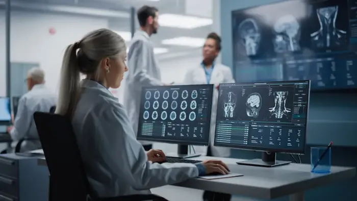
Your Guide to Radiology: Understanding Scans, Safety, and More
Explore our comprehensive radiology guide to understand X-rays, CT, MRI, ultrasound, safety tips, and what to expect from scans for accurate diagnosis and care.

Written by Dr. Md Yusuf Shareef
Reviewed by Dr. Dhankecha Mayank Dineshbhai MBBS
Last updated on 13th Jan, 2026

Introduction
When your doctor says, "We need to get a scan," it can be a moment of uncertainty. What exactly is an MRI? How is a CT scan different? Is the radiation safe? The field of radiology, dedicated to imaging the human body, is central to modern medicine, yet it often feels like a mystery to patients. This guide will demystify the world of medical imaging, explaining the different types of scans, their purposes, what you can expect, and how they help your doctor provide the best possible care. We’ll cover everything from common X-rays to advanced interventional procedures, ensuring you feel informed and confident.
What is Radiology? The Science of Seeing Inside the Body
Radiology is the medical discipline that uses imaging technology to diagnose, monitor, and sometimes even treat diseases. It allows physicians to see inside the body without the need for surgery, providing critical information about the structure and function of organs, bones, and tissues. The journey of radiology began with Wilhelm Conrad Röntgen's discovery of X-rays in 1895, a breakthrough that earned him the first Nobel Prize in Physics. Since then, the field has exploded with innovations like ultrasound, CT, and MRI, making it an indispensable tool for nearly every medical specialty, from oncology to orthopedics.
The Vital Role of a Radiologist
A common misconception is that the technologist who operates the scanning machine is the one interpreting the results. In reality, that crucial task falls to a radiologist. These are medical doctors who have completed specialised training in interpreting medical images. They act as expert consultants to your primary physician, providing a detailed report that aids in diagnosis. Their expertise is not just in operating the machinery but in discerning subtle abnormalities, correlating imaging findings with clinical symptoms, and recommending the next appropriate steps.
Consult a Radiologist for the best advice
Major Types of Diagnostic Imaging Techniques
Each imaging technique offers a unique window into the body. The choice of scan depends on what part of the body is being examined and what information the doctor needs.
X-Rays: The First Revolution in Medical Imaging
X-rays are a form of electromagnetic radiation that can pass through most objects, including the body. Dense materials like bone absorb more radiation and appear white on the resulting image, while softer tissues appear in shades of gray. X-rays are incredibly fast, inexpensive, and excellent for evaluating:
1. Broken bones (fractures)
2. Dental problems
3. Chest infections (like pneumonia)
4. Breast tissue (in a specialised form called a mammogram)
Computed Tomography (CT) Scans: Detailed Cross-Sectional Views
A CT scan (or "CAT scan") uses a series of X-ray beams taken from different angles to create cross-sectional images, or "slices," of the body. A computer then assembles these slices into highly detailed 2D and 3D images. Think of it as looking at a loaf of bread by examining each slice rather than just the whole loaf from the outside. CT scans are invaluable for:
1. Detecting internal bleeding and injuries from trauma
2. Diagnosing cancers and monitoring their response to treatment
3. Guiding biopsies and other minimally invasive procedures
4. Visualising complex bone fractures
Magnetic Resonance Imaging (MRI): Powerful Images Without Radiation
Unlike X-rays and CT scans, an MRI uses a powerful magnet and radio waves to generate detailed images of organs and soft tissues. It does not involve ionising radiation. The MRI is exceptionally good at distinguishing between normal and abnormal tissue, making it the preferred choice for examining:
1. The brain and spinal cord
2. Joints, muscles, and ligaments
3. The heart and blood vessels
4. Organs in the pelvis and abdomen
A key point of confusion for many is the difference between an MRI and a CT scan. While both provide detailed images, CT is generally better for bone and lung imaging, and is much faster. MRI provides superior soft tissue contrast and is essential for neurological and musculoskeletal disorders.
Ultrasound: Safe Imaging Using Sound Waves
Ultrasound imaging, or sonography, uses high-frequency sound waves to produce real-time images of the inside of the body. It is completely radiation-free, which makes it the go-to method for monitoring fetal development during pregnancy. It is also widely used to examine:
1. Abdominal organs (liver, kidneys, gallbladder)
2. The heart (echocardiogram)
3. Blood flow (Doppler ultrasound)
4. Muscles and tendons
Nuclear Medicine (PET & SPECT Scans)
This branch of radiology involves administering a small amount of radioactive material (a tracer) into the patient's body. This tracer accumulates in specific organs or tissues. Special cameras then detect the radiation emitted and create images that show how those organs are functioning, not just what they look like. A common example is the PET scan, which is crucial in oncology for detecting cancer spread (metastasis) and evaluating brain disorders.
Interventional Radiology: Diagnosis and Treatment in One
Interventional radiology (IR) is a fascinating sub-specialty where radiologists use imaging guidance (like X-ray or ultrasound) to perform minimally invasive procedures. Instead of large incisions, they use tiny needles and catheters threaded through blood vessels or other pathways. This "pinhole surgery" approach offers faster recovery, less pain, and lower risk than traditional surgery. Common interventional radiology procedures include:
1. Angioplasty and Stenting: Opening blocked arteries.
2. Embolisation: Blocking blood flow to tumors or to stop bleeding.
3. Ablation: Using heat or cold to destroy tumors.
4. Needle Biopsies: Precisely extracting tissue samples for diagnosis.
What to Expect During Your Radiology Procedure
Knowing what to expect can ease anxiety. While each test is different, a general process applies.
How to Prepare for Common Scans
Here are some simple steps to help you prepare for different types of common scans:
• MRI/CT: You may be asked to avoid eating or drinking for a few hours beforehand. You will need to remove all metal objects. For an MRI, you must inform the staff if you have any metal implants. If you experience claustrophobia, your doctor can discuss options like an "open MRI" or a mild sedative.
• Ultrasound: Preparation might involve drinking water to fill your bladder (for pelvic ultrasounds) or fasting (for abdominal ultrasounds).
• X-Ray: Typically, no special preparation is needed.
If your condition requires a scan with contrast dye and you have underlying kidney issues, your doctor may recommend a blood test beforehand to ensure it's safe. Apollo24|7 offers convenient home collection for tests like creatinine clearance to simplify this process.
Understanding Your Radiology Report
After the scan, the radiologist will analyse the images and send a detailed report to your referring doctor. This report describes what was seen, notes any abnormalities, and provides an impression or diagnosis. It's important to discuss the results with your doctor, who can interpret the findings in the context of your overall health. They can explain what the technical terms mean for your specific situation.
Safety in Radiology: Radiation, Contrast Dye, and FAQs
Safety is a paramount concern. The radiation dose from standard X-rays is very low, comparable to the natural background radiation we're exposed to over a few days. CT scans involve higher doses, but the diagnostic benefit almost always outweighs the minimal long-term risk. Radiologists follow the "ALARA" principle—As Low As Reasonably Achievable—to minimise exposure.
Contrast dye, used in some CTs, MRIs, and other procedures, is generally safe but can cause allergic reactions in a small percentage of people. Be sure to inform your healthcare team of any allergies or kidney problems. If you experience any unusual symptoms after a scan, such as a persistent rash or swelling, consult a doctor online with Apollo24|7 for immediate advice.
The Future of Radiology: AI and Advanced Imaging
The future of radiology is intelligent and precise. Artificial intelligence (AI) is already being used to help radiologists detect subtle patterns in images, such as early signs of cancer in mammograms, potentially improving accuracy and speed. Advanced techniques like molecular imaging aim to detect diseases at the cellular level long before structural changes occur. This continuous innovation promises even earlier diagnoses and more personalised treatment plans.
Conclusion
Understanding radiology empowers you to be an active participant in your healthcare journey. From the simple X-ray that confirms a fracture to the sophisticated MRI that maps a brain tumor, these imaging tools are fundamental to modern medicine. They reduce uncertainty, guide effective treatment, and save lives. While the technology can seem intimidating, knowing the basics of how each scan works and what to expect can transform anxiety into confidence. Remember, these procedures are performed by highly skilled professionals dedicated to your well-being. Always maintain open communication with your doctor, ask questions about recommended scans, and discuss the results thoroughly. If your doctor has recommended a scan and you have further questions about the procedure or its necessity, booking a consultation with a specialist through a platform like Apollo24|7 can provide the clarity you need.
Consult a Radiologist for the best advice
Consult a Radiologist for the best advice

Dr Suchana Kushvaha
Radiologist
14 Years • MBBS, MD Radiodiagnosis/Radiology, FNB ( Breast Imaging)
Gurugram
Prajnam Complete Breast Care, Gurugram

Dr.priyank Ks Chaudhary
Radiologist
10 Years • MBBS DMRD & DNB
Lucknow
Apollo Clinic Hazratganj, Lucknow
Dr Manik Bandyopadhyay
Radiologist
44 Years • MBBS, DMR
Kolkata
RHCC - Rejuvenecer Healthcare Centre, Kolkata

Dr. Surendra K
Radiologist
29 Years • MBBS, MD (Radio-Diagnosis)
Bengaluru
Apollo Clinic, Koramangala, Bengaluru
Dr.kiran Barla
Radiologist
5 Years • MBBS, DNB (Radiology)
Hanamkonda
Apollo Clinic Hanamkonda, Hanamkonda
Consult a Radiologist for the best advice

Dr Suchana Kushvaha
Radiologist
14 Years • MBBS, MD Radiodiagnosis/Radiology, FNB ( Breast Imaging)
Gurugram
Prajnam Complete Breast Care, Gurugram

Dr.priyank Ks Chaudhary
Radiologist
10 Years • MBBS DMRD & DNB
Lucknow
Apollo Clinic Hazratganj, Lucknow
Dr Manik Bandyopadhyay
Radiologist
44 Years • MBBS, DMR
Kolkata
RHCC - Rejuvenecer Healthcare Centre, Kolkata

Dr. Surendra K
Radiologist
29 Years • MBBS, MD (Radio-Diagnosis)
Bengaluru
Apollo Clinic, Koramangala, Bengaluru
Dr.kiran Barla
Radiologist
5 Years • MBBS, DNB (Radiology)
Hanamkonda
Apollo Clinic Hanamkonda, Hanamkonda
More articles from General Medical Consultation
Frequently Asked Questions
1. How long does it take to get radiology results?
The time varies. For urgent cases in an emergency room, results can be available within an hour. For routine outpatient scans, it typically takes 24 to 48 hours for the radiologist to interpret the images and send a report to your doctor.
2. What is the difference between a radiologist and a radiographer?
A radiologist is a medical doctor who specialises in interpreting medical images. A radiographer (or radiology technologist) is the healthcare professional who is trained to operate the imaging equipment and perform the scan safely.
3. Are there any risks associated with repeated MRI scans?
An MRI does not use ionising radiation and is considered very safe. There are no known long-term risks from the magnetic fields and radio waves used. The main considerations are for people with certain metal implants or devices.
4. Can I get a CT scan if I am pregnant?
As a precaution, CT scans are generally avoided during pregnancy, especially of the abdomen and pelvis, due to the radiation exposure to the fetus. If a scan is medically essential, the radiologist will use the lowest possible dose to get the necessary information. Ultrasound or MRI is often a preferred alternative during pregnancy.
5. Why do I need a contrast dye for my scan?
Contrast dye (or 'contrast agent') is used to highlight specific blood vessels, organs, or tissues, making them easier to see and distinguish from surrounding structures. This greatly improves the diagnostic accuracy of the scan.


_0.webp)

