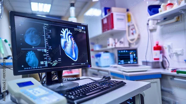
Angiogram: Purpose And Procedure Of Angiogram
Learn about the purpose of an angiogram and the procedure involved. Understand how this diagnostic test helps detect blockages in blood vessels and aids in the treatment of heart and vascular conditions.

Written by Dr Shreya Sarkar
Last updated on 3rd Jul, 2025
Heart diseases are a leading cause of mortality worldwide, and early diagnosis plays a critical role in effective treatment and management. One of the pivotal diagnostic tools in cardiology is the angiogram. This medical imaging technique offers doctors a detailed view of blood vessels and helps them evaluate blood flow and detect blockages, narrowing, or abnormalities. Whether you're dealing with chest pain, shortness of breath, or unexplained fatigue, your doctor may recommend an angiogram to understand what might be going on inside your arteries.
This article will provide a comprehensive overview of angiograms, including their purpose, procedure, preparation, recovery, and what patients can expect during the process. Understanding the angiogram will not only help you feel more confident about the procedure but also empower you with valuable knowledge about your heart health.
What is an Angiogram?
An angiogram, also known as coronary angiography when involving the heart, is a medical imaging test used to visualise blood vessels. During the procedure, a special contrast dye is injected into the blood vessels, making them visible on X-ray images. These images help doctors evaluate the condition of the arteries and identify any areas that might be blocked, narrowed, or damaged. An angiogram is often considered the "gold standard" for diagnosing coronary artery disease (CAD) and other cardiovascular conditions.
While angiograms can be performed on various parts of the body, the most common use is to examine the coronary arteries of the heart. This procedure can help identify conditions such as:
Coronary artery disease (CAD): A condition where plaque builds up in the coronary arteries, potentially leading to a heart attack.
Aneurysms: Weak areas in the arteries that can cause them to bulge.
Arterial blockages: Obstructions in the arteries that can restrict blood flow, increasing the risk of heart attacks or strokes.
Why is an Angiogram Performed?
An angiogram serves several purposes, primarily focused on diagnosing heart disease, evaluating blood flow, and determining appropriate treatment options. Some common reasons your doctor might recommend an angiogram include:
Diagnosis of Coronary Artery Disease (CAD): CAD is one of the most common heart conditions caused by the buildup of plaque in the coronary arteries. An angiogram allows your doctor to see the location and severity of any blockages, which can help them determine the best course of action.
Pre-surgical Planning: Before performing certain surgeries, such as coronary artery bypass grafting (CABG) or angioplasty, an angiogram provides crucial information about the condition of the blood vessels to guide treatment decisions.
Evaluation of Heart Function: If you’ve been experiencing symptoms such as chest pain, shortness of breath, or fatigue, an angiogram can help pinpoint the cause by revealing potential problems with the arteries.
Assessment of Stents or Bypass Grafts: After you've had procedures like stent placement or heart bypass surgery, an angiogram may be performed to ensure that the treated areas have good blood flow and are functioning properly.
Detecting Aneurysms or Other Vascular Abnormalities: Angiograms are also used to identify aneurysms, malformations, or other issues in the arteries that could impact blood flow.
How is an Angiogram Performed?
An angiogram is typically performed in a hospital's catheterisation lab, also known as a "cath lab." It is considered a minimally invasive procedure that involves the use of a catheter (a thin, flexible tube) to deliver a contrast dye into the arteries. Here’s a step-by-step breakdown of how the angiogram procedure works:
1. Preparation
Before the procedure, you will likely be asked to fast for several hours. This is important to prevent any complications with anaesthesia or sedation during the angiogram. Your healthcare team will provide specific instructions, and it's essential to follow them carefully.
Health History: Your doctor will ask about any allergies (especially to iodine or contrast dyes) and medical conditions you have, including kidney disease, diabetes, or blood clotting disorders.
Pre-procedure tests: You may undergo a physical examination, blood tests, or an electrocardiogram (ECG) to assess your heart function.
2. Sedation
An angiogram is generally performed under local anaesthesia to numb the area where the catheter will be inserted, typically the groin or wrist. In some cases, mild sedation may also be provided to help you relax. However, the procedure is usually not painful, and many patients remain awake and alert.
3. Insertion of the Catheter
Once the area is numbed, the doctor will insert a small needle to create a tiny opening in the skin. They will then insert the catheter into the blood vessel. Using live X-ray imaging (fluoroscopy), the doctor guides the catheter through the bloodstream toward the area that needs to be examined, such as the coronary arteries.
4. Injection of Contrast Dye
Once the catheter is in place, a contrast dye will be injected into the blood vessels. The dye makes the blood vessels visible on the X-ray images, allowing the doctor to see any blockages, narrowing, or abnormalities in the arteries. You may feel a warm sensation when the dye is injected, which is normal.
5. Imaging and Evaluation
The doctor will collect a series of X-ray images while the dye moves through your blood vessels. These images will be analysed to assess the flow of blood and identify any issues with the arteries. In some cases, additional images or tests may be required for a more detailed examination.
6. Completion of the Procedure
Once the images have been taken and the doctor has evaluated the results, the catheter will be carefully removed, and the tiny opening in your skin will be closed. If the catheter was inserted through the wrist, a bandage or pressure dressing may be applied to the site.
7. Recovery
After the procedure, you will be monitored for a short time in a recovery area. The duration of the recovery period may vary depending on the type of procedure and your overall health. You would be advised to stay still for a few hours if the catheter was inserted into the groin to allow the puncture site to heal. If the wrist is used, you can move around sooner.
Risks and Complications
While angiograms are generally safe, like all medical procedures, there are some risks involved. These may include:
Allergic reaction: Some people may experience an allergic reaction to the contrast dye, though this is rare.
Bleeding or infection: Any invasive procedure carries a risk of bleeding or infection at the catheter insertion site.
Damage to blood vessels: In very rare cases, the catheter may cause damage to the blood vessels.
Kidney problems: The contrast dye can sometimes affect kidney function, especially in patients with pre-existing kidney conditions.
It’s important to discuss these risks with your healthcare provider before the procedure.
What to Expect After the Angiogram
Following the angiogram, you may experience mild soreness or bruising at the site where the catheter was inserted. This is normal and should resolve on its own. In some cases, you may be given specific instructions about when you can resume normal activities, such as driving or exercise.
If the angiogram was done to evaluate blockages or narrowing, your doctor may recommend further treatments, such as:
Angioplasty: A procedure in which a small balloon is inflated inside the blocked artery to open it up.
Stent placement: A small mesh tube is inserted to keep the artery open.
Medications: Your doctor may prescribe medications to manage risk factors, such as cholesterol-lowering drugs or blood thinners.
Conclusion
An angiogram is a valuable tool for diagnosing heart disease and other vascular conditions. By providing clear, detailed images of blood vessels, it allows doctors to identify blockages, aneurysms, and other issues that may be affecting blood flow. Though the procedure may sound intimidating, it is minimally invasive, typically well-tolerated, and an essential step in determining the most effective treatment for your heart health.
If you're about to undergo an angiogram, it’s important to ask your healthcare provider any questions you may have about the procedure, potential risks, and what to expect during recovery. By staying informed and prepared, you can approach the procedure with confidence and take an active role in your heart health journey.
Consult Top Cardiologists
Consult Top Cardiologists

Dr. Tripti Deb
Cardiologist
40 Years • MBBS, MD, DM, FACC, FESC
Hyderabad
Apollo Hospitals Jubilee Hills, Hyderabad
Dr Moytree Baruah
Cardiologist
10 Years • MBBS, PGDCC
Guwahati
Apollo Clinic Guwahati, Assam, Guwahati

Dr. Zulkarnain
General Physician
2 Years • MBBS, PGDM, FFM
Bengaluru
PRESTIGE SHANTHINIKETAN - SOCIETY CLINIC, Bengaluru

Dr. Janjirala Seshivardhan
Cardiologist
7 Years • MBBS,DNB(GM),DM(Cardiology)
Manikonda Jagir
Apollo Clinic, Manikonda, Manikonda Jagir

Dr. Anand Ravi
General Physician
2 Years • MBBS
Bengaluru
PRESTIGE SHANTHINIKETAN - SOCIETY CLINIC, Bengaluru


