Atelectasis and Breathlessness: Causes, Symptoms, Diagnosis & Treatment
Learn about atelectasis, its causes, symptoms, and how it leads to breathlessness. Discover diagnosis, treatment, and prevention strategies for better lung health.


Introduction
Have you ever felt short of breath and wondered what might be causing it? Breathlessness can be unsettling, especially when it occurs suddenly. One possible reason behind this discomfort is atelectasis, a condition that affects the lungs and can make breathing difficult. It is a lung condition where one or more sections of your lungs fail to expand properly or collapse entirely. This reduces the amount of oxygen entering your bloodstream and makes it harder for the body to function efficiently.
Breathlessness, or dyspnoea, is the sensation of not being able to breathe properly. It can range from mild discomfort to severe difficulty in drawing air into the lungs. Various factors, such as lung diseases, infections, heart problems, or anxiety, can make you breathless. In the case of atelectasis, the lack of proper lung expansion may lead to inadequate oxygen exchange which can make your breathing more laboured.
Understanding the causes and symptoms of atelectasis can help you take the right steps toward better lung health. Let’s explore what this condition is and how it contributes to breathlessness.
Consult Top Pulmonologist
Understanding Atelectasis
Atelectasis directly impacts how well your lungs function. When parts of your lungs collapse, they can’t take in oxygen efficiently, which can leave you feeling breathless or fatigued. This condition can be temporary or long-term, depending on its cause.
1. Types of Atelectasis
Atelectasis can be classified into different types based on its underlying cause:
Obstructive Atelectasis – This occurs when something blocks the airway, such as mucus, a tumour, or an inhaled foreign object. The blocked portion of the lung collapses as air is absorbed but not replaced.
Non-Obstructive Atelectasis – This happens due to factors other than blockages, such as compression, where external pressure (from fluid, tumours, or enlarged organs) prevents the lung from expanding. It can also result from postoperative changes, where shallow breathing after surgery leads to lung collapse.
Adhesive Atelectasis – This occurs when a lack of surfactant, a substance that helps keep the lungs open, causes the lung tissues to stick together. It’s common in premature babies or those with lung infections.
Cicatricial Atelectasis – This type develops due to lung scarring from infections, radiation, or chronic lung disease, making the lung tissue shrink and collapse.
2. How Atelectasis Affects Breathing?
When part of the lung collapses, it reduces the amount of oxygen your body receives. This can lead to shallow breathing, rapid breaths, and a feeling of tightness in the chest. The lungs rely on full expansion to function properly, so when air can’t reach certain areas, you may feel breathless, tired, or even dizzy.
Mild atelectasis may not cause noticeable symptoms, but in severe cases, it can lead to low oxygen levels, making daily activities harder. If left untreated, it can increase the risk of infections like pneumonia.
Causes of Atelectasis
Atelectasis can develop for many reasons, but the underlying cause usually falls into two main categories: obstructive and non-obstructive. Understanding what triggers lung collapse can help in early detection and management.
1. Obstructive Causes
Obstructive atelectasis happens when something blocks the airway, preventing air from reaching the lungs. As a result, the trapped air gets absorbed, and the affected lung section collapses. Common causes include:
Mucus plugs – Thick mucus can build up in the airways, often after surgery or due to lung conditions like cystic fibrosis.
Tumours – A growth inside the airway can restrict airflow, gradually leading to lung collapse.
Foreign objects – Inhaled food, small objects, or other substances can block the airways, especially in children.
2. Non-Obstructive Causes
Non-obstructive atelectasis occurs without a physical blockage but still leads to lung collapse due to external factors. These may include:
Compression – Fluid build-up (pleural effusion), enlarged organs, or tumours can press against the lungs, preventing full expansion.
Post-surgical effects – Shallow breathing due to pain after surgery can cause lung collapse, particularly in those undergoing chest or abdominal procedures.
Surfactant deficiency – A lack of surfactant, a substance that prevents lung tissues from sticking together, can lead to atelectasis, commonly seen in premature babies.
Lung scarring (fibrosis) – Previous infections, radiation, or chronic lung diseases can cause lung tissue to shrink, reducing its ability to expand.
Both types of atelectasis can affect breathing and oxygen levels, making early diagnosis and treatment essential.
Causes of Breathlessness
Breathlessness can arise from various medical conditions. While occasional shortness of breath may not be concerning, persistent or sudden breathlessness could indicate an underlying health issue. The causes generally fall into two main categories: respiratory system disorders and cardiovascular conditions.
1. Respiratory System Disorders
Some of the common respiratory causes of breathlessness include:
Atelectasis – When parts of the lung collapse, it reduces oxygen intake, causing shallow or laboured breathing.
Chronic Obstructive Pulmonary Disease (COPD) – A progressive lung condition that leads to airway narrowing and difficulty exhaling.
Asthma – Airways become inflamed and constricted, causing wheezing and shortness of breath.
Pneumonia – Infection in the lungs leads to fluid accumulation, making breathing more difficult.
Pulmonary fibrosis – Scarring in the lungs reduces elasticity, limiting lung expansion and oxygen exchange.
2. Cardiovascular Conditions
The heart and lungs work together to deliver oxygen throughout the body. When the heart is compromised, it can lead to breathlessness. Some common cardiovascular causes include:
Heart failure – The heart struggles to pump blood efficiently, causing fluid build-up in the lungs and shortness of breath.
Coronary artery disease – Narrowed arteries restrict blood flow to the heart, leading to breathlessness during physical activity.
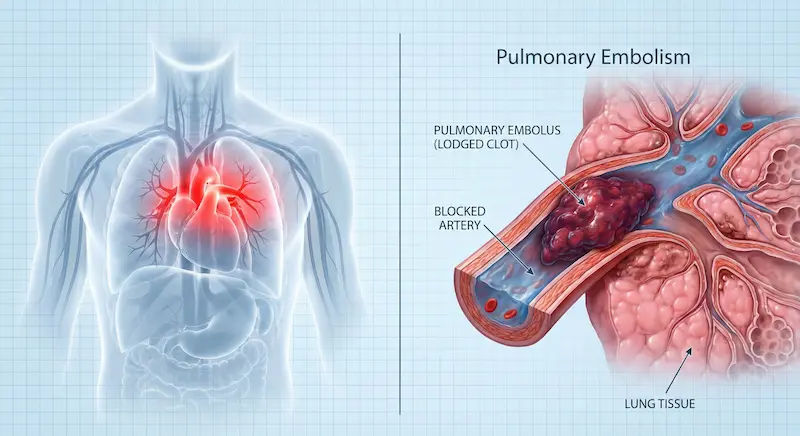
Pulmonary embolism – A blood clot in the lungs reduces oxygen levels, causing sudden and severe shortness of breath.
Arrhythmias – Irregular heartbeats can affect blood circulation, making it harder for the body to get enough oxygen.
Relationship Between Atelectasis and Breathlessness
Atelectasis and breathlessness are closely linked because the lungs play a vital role in oxygen exchange. When a part of your lung collapses, your body struggles to get enough oxygen which leads to shortness of breath.
1. Pathophysiology
Atelectasis reduces lung volume, decreasing oxygen intake and leading to hypoxia (low oxygen levels). This triggers an increase in respiratory rate as the body tries to compensate. The severity of breathlessness depends on the extent of lung collapse and the individual’s overall lung function.
2. Compensatory Mechanisms
The body responds to atelectasis by increasing breathing rate and heart rate to maintain oxygen delivery. Nearby lung regions may try to expand more to compensate for the affected area. Over time, however, untreated atelectasis can lead to worsening breathlessness and an increased risk of infections.
Risk Factors for Atelectasis and Associated Breathlessness
Atelectasis can develop due to a variety of factors that interfere with normal lung function. Some risk factors are related to underlying medical conditions, while others are associated with surgical procedures or prolonged immobility. Understanding these risk factors can help in early detection and prevention.
1. Medical Conditions
Certain health conditions weaken the lungs or obstruct airways, increasing the likelihood of atelectasis. These include:
Chronic lung diseases: Conditions such as COPD and asthma lead to airway narrowing and mucus build-up.
Neuromuscular disorders: These conditions affect the muscles needed for deep breathing, making lung expansion difficult.
Obesity: Excess body weight increases pressure on the lungs and reduces the ability to take deep breaths.
Prolonged immobility: leads to shallow breathing and mucus accumulation, especially in bedridden patients.
2. Surgical Procedures
Postoperative atelectasis is common, particularly after major surgeries. Some factors that contribute to lung collapse include:
Chest or abdominal surgery: The pain caused by these procedures affects deep breathing, leading to reduced lung expansion.
General anaesthesia: It temporarily suppresses normal breathing patterns and lung function.
Prolonged intubation: This can cause mucus accumulation and airway blockages, increasing the risk of lung collapse.
Symptoms and Clinical Presentation
The symptoms of atelectasis vary depending on how much of the lung is affected. In mild cases, a person may not notice any symptoms, but when a larger portion of the lung collapses, breathing difficulties become more apparent. The body struggles to get enough oxygen, leading to noticeable respiratory and systemic signs.
1. Respiratory Symptoms
Atelectasis directly impacts lung function, making it harder to breathe. Some common respiratory symptoms include:
Shortness of breath: This worsens with physical activity as the lungs struggle to meet oxygen demands.
Shallow or rapid breathing: It is due to reduced lung expansion which makes each breath feel less effective.
Persistent coughing: It occurs as the body tries to clear mucus from blocked or collapsed areas of the lung.
Chest discomfort or tightness: This is caused by reduced lung volume and pressure changes within the chest cavity.
2. Systemic Signs
When oxygen levels drop due to atelectasis, the body reacts in several ways. Systemic symptoms may include:
Cyanosis (bluish tint on lips or skin): It is due to insufficient oxygen reaching the bloodstream.
Fatigue and dizziness: Caused by inadequate oxygen supply which leads to reduced energy levels and lightheadedness.
Increased heart rate (tachycardia): It is a biological response that occurs when the body tries to compensate for low oxygen by pumping blood faster.
Fever: It develops due to mucus build-up in the collapsed lung area, especially due to an infection such as pneumonia.
3. Diagnostic Approach
Since atelectasis may not always present with obvious symptoms, diagnostic tests play a crucial role in confirming lung collapse and identifying its underlying cause. Doctors rely on imaging and pulmonary function tests to assess lung function and determine the severity of the condition.
4. Imaging Studies
Medical imaging is the most effective way to detect atelectasis. Common imaging tests include:
Chest X-ray: Provides a quick and simple way to identify collapsed lung areas.
CT scan: Gives a more detailed view of the lungs, especially for identifying blockages or underlying lung diseases.
Ultrasound: Helps detect fluid accumulation that may be compressing the lungs.
5. Pulmonary Function Tests
Lung function tests help measure how well the lungs are working. These tests include:
Spirometry: Evaluates lung capacity and airflow to help detect restricted breathing patterns.
Oxygen saturation (pulse oximetry): Measures how much oxygen is in the blood, indicating how well the lungs are functioning.
Arterial blood gas (ABG) test: Checks oxygen and carbon dioxide levels in the blood, helping to assess the severity of respiratory impairment.
Management Strategies
The goal of treating atelectasis is to reopen the collapsed lung areas, clear airway obstructions, and improve oxygen levels. Depending on the cause and severity of the condition, treatment may involve medical interventions or, in some cases, surgical procedures.
1. Medical Treatments
Non-invasive treatments are the first approach to managing atelectasis. These include:
Breathing exercises (incentive spirometry): Support deep breathing to expand the lungs and prevent further collapse.
Oxygen therapy: Helps improve oxygen levels in cases of severe atelectasis.
Mucus-clearing techniques( such as chest physiotherapy, postural drainage, or suctioning): Help in removing blockages from the airways.
Bronchodilators and corticosteroids: Used in patients with underlying respiratory conditions like asthma or COPD to relax and open the airways.
2. Surgical Interventions
If conservative treatments are not effective, surgical procedures may be necessary. These include:
Bronchoscopy: It is a procedure that removes mucus plugs, foreign objects, or tumours that may be blocking the airways.
Thoracentesis: Drains excess fluid from around the lungs to relieve compression and allow normal lung expansion.
Lung surgery: Required in severe cases where lung damage is irreversible, such as in cases of advanced fibrosis or tumours.
Preventive Measures
Atelectasis is often preventable, especially in patients undergoing surgery or those with underlying lung conditions. Preventive strategies focus on maintaining lung function, improving breathing patterns, and reducing airway blockages.
1. Postoperative Care
Patients recovering from surgery are at higher risk of developing atelectasis, so post-surgical lung care is crucial. Preventive measures include:
Deep breathing exercises: help re-expand the lungs and prevent mucus accumulation.
Early mobilisation: encourages movement to reduce the risk of lung collapse and improve circulation.
Pain management: allows patients to take deeper breaths without discomfort, reducing the likelihood of shallow breathing.
2. Lifestyle Modifications
Making simple lifestyle changes can help maintain healthy lung function and lower the risk of atelectasis. These include:
Quitting smoking: reduces mucus build-up and prevents lung damage.
Staying physically active: promotes lung expansion and keeps the respiratory system strong.
Staying hydrated: helps thin mucus, making it easier to clear from the airways.
Maintaining good posture: especially when sitting for long periods, to allow full lung expansion and prevent lung compression.
By following these preventive strategies, individuals can significantly reduce their risk of developing atelectasis and related breathing difficulties.
Consult Top Pulmonologist
Consult Top Pulmonologist

Dr. Bichitra Ojha
Pulmonology Respiratory Medicine Specialist
9 Years • MBBS, MD( Respiratory Medicine)
Kolkata
VDC Clinic, Kolkata

Dr. Tamal Bhattacharyya
Pulmonology Respiratory Medicine Specialist
8 Years • MBBS, MD (Respiratory Medicine)
Kolkata
MCR SUPER SPECIALITY POLY CLINIC & PATHOLOGY, Kolkata
Dr. Preeti Kathail
General Physician/ Internal Medicine Specialist
17 Years • MBBS, PGDHHM
Bangalore
Apollo Clinic Bellandur, Bangalore

Dr Sravani Kuppam
General Physician/ Internal Medicine Specialist
12 Years • MBBS DNB General Medicine, CCDM (Diabetes)
Bengaluru
Apollo Medical Center, Marathahalli, Bengaluru

Dr Vishwa Vijeth K.
Pulmonology Respiratory Medicine Specialist
8 Years • MBBS, MD ( Respiratory Medicine)
Bangalore
Apollo Clinic Bellandur, Bangalore
Consult Top Pulmonologist
Dr. Bichitra Ojha
Pulmonology Respiratory Medicine Specialist
9 Years • MBBS, MD( Respiratory Medicine)
Kolkata
VDC Clinic, Kolkata
Dr. Tamal Bhattacharyya
Pulmonology Respiratory Medicine Specialist
8 Years • MBBS, MD (Respiratory Medicine)
Kolkata
MCR SUPER SPECIALITY POLY CLINIC & PATHOLOGY, Kolkata
Dr. Preeti Kathail
General Physician/ Internal Medicine Specialist
17 Years • MBBS, PGDHHM
Bangalore
Apollo Clinic Bellandur, Bangalore
Dr Sravani Kuppam
General Physician/ Internal Medicine Specialist
12 Years • MBBS DNB General Medicine, CCDM (Diabetes)
Bengaluru
Apollo Medical Center, Marathahalli, Bengaluru
Dr Vishwa Vijeth K.
Pulmonology Respiratory Medicine Specialist
8 Years • MBBS, MD ( Respiratory Medicine)
Bangalore
Apollo Clinic Bellandur, Bangalore
.webp)
.webp)