
Sialendoscopy: A Minimally Invasive Solution for Salivary Gland Stones
Discover how sialendoscopy offers a minimally invasive solution for salivary gland stones. Learn about the procedure, benefits, recovery, and how it can relieve pain and restore normal salivary flow.

Written by Dr. Siri Nallapu
Reviewed by Dr. D Bhanu Prakash MBBS, AFIH, Advanced certificate in critical care medicine, Fellowship in critical care medicine
Last updated on 13th Jan, 2026

Introduction
Imagine the frustrating pain of a swollen cheek every time you eat, a constant, dull ache that flares up with every meal. For many, this isn't just an inconvenience; it's the tell-tale sign of a blocked salivary gland, often caused by tiny stones. Traditionally, treating these obstructions involved invasive surgery, external incisions, and significant recovery time. But medical science has made a leap forward. Enter sialendoscopy, a groundbreaking minimally invasive procedure that has revolutionised the diagnosis and treatment of salivary gland disorders. This guide will walk you through everything you need to know about sialendoscopy—from what causes these painful blockages and the symptoms to watch for, to a detailed look at the procedure itself, its immense benefits, and what recovery truly looks like. If you're seeking a modern solution that prioritises gland preservation and your comfort, you're in the right place.
What is a Salivary Gland Obstruction?
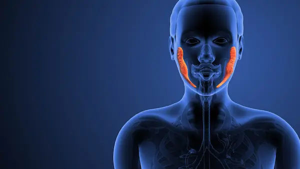
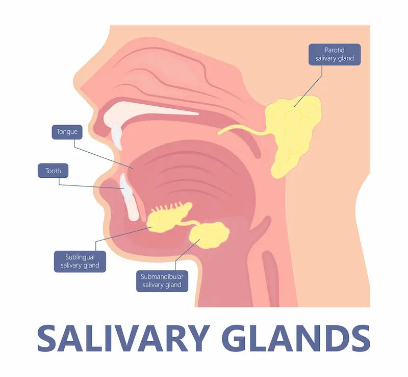
Your salivary glands (the major ones being the parotid, submandibular, and sublingual glands) produce saliva, which is essential for digestion, swallowing, and oral health. Sometimes, the ducts that carry this saliva into your mouth can become blocked. This obstruction prevents saliva from draining, leading to a painful backup and swelling, particularly during meals when saliva production increases.
Common Causes of Blocked Salivary Glands
Salivary Stones (Sialolithiasis)
The most common culprit is salivary stones, or sialoliths. These are calcified structures that form from the minerals in saliva, much like kidney stones. They can vary in size from a grain of sand to several centimeters. Stones occur most frequently in the submandibular glands due to the thicker, mucus-rich saliva and the upward course of its duct.
Strictures and Scar Tissue
Narrowing of the salivary ducts, known as strictures, can also cause blockages. These can result from previous inflammation, injury, or infection. The narrow passage makes it easy for saliva to get stuck, even without a stone.
Mucous Plugs and Debris
In some cases, thick mucus or cellular debris can clump together, forming a plug that obstructs the duct. This is often associated with dehydration or certain medical conditions.
Consult an ENT specialist for the best advice
What is Sialendoscopy? The Modern Diagnostic and Therapeutic Tool
Sialendoscopy is a state-of-the-art, minimally invasive technique that allows an ENT surgeon to visually examine the inside of the salivary ducts, diagnose the exact cause of a blockage, and treat it simultaneously. Think of it as a tiny, high-tech camera journey into the intricate plumbing of your salivary system.
How Does Sialendoscopy Work? A Look at the Technology
The procedure utilises an ultra-thin endoscope—a flexible or semi-rigid tube less than 1mm in diameter equipped with a light, a camera, and a working channel. This micro-instrument is gently inserted into the natural opening of the salivary duct in the mouth. The camera sends real-time images to a monitor, giving the surgeon a magnified view of the duct's interior. Through the working channel, miniature surgical instruments like baskets, forceps, balloons, and lasers can be passed to remove stones, dilate strictures, or flush out debris.
Are You a Candidate? Symptoms of Salivary Gland Blockage
How do you know if you might need a sialendoscopy procedure? The symptoms are often unmistakable and meal-related:
Painful swelling in the cheek or under the jaw, especially when eating or drinking.
A tender, firm lump that you can feel.
Recurrent infections in the gland (sialadenitis), characterised by increased pain, redness of the skin over the gland, and sometimes fever or pus draining into the mouth.
Persistent dry mouth on one side.
If these symptoms of a blocked salivary gland persist beyond two weeks or keep recurring, it is crucial to consult a specialist. You can consult an ENT specialist online with Apollo24|7 for an initial evaluation and to discuss if a physical visit is necessary.
The Sialendoscopy Procedure: What to Expect Step-by-Step
Step 1: Diagnosis and Pre-Procedure Imaging
Your doctor will first confirm the obstruction using imaging. This may include an ultrasound or a non-contrast CT scan, which are excellent for detecting stones. Sometimes, a conventional X-ray (sialogram) might be used.
Step 2: The Day of the Procedure – Anesthesia and Setup
Sialendoscopy is typically performed as an outpatient procedure under local or general anesthesia, depending on the complexity and patient comfort. You will be able to go home the same day.
Step 3: The Intricate Process – Navigation, Inspection, and Treatment
The surgeon will dilate the natural duct opening in the mouth before guiding the sialendoscope inside. They will then:
Navigate through the ductal system, inspecting the walls for stones, strictures, or inflammation.
Treat the problem: A stone is captured in a tiny basket or fragmented with a laser; a stricture is dilated with a microscopic balloon.
Flush the duct with saline to clear any remaining particles.
Step 4: Post-Procedure Care and Immediate Recovery
After the scope is removed, you will spend a short time in recovery. You might be asked to suck on sour candies to stimulate saliva flow and keep the ducts open. Most patients experience minimal discomfort.
Benefits of Choosing Sialendoscopy Over Traditional Surgery
The advantages of this minimally invasive salivary stone removal technique are profound:
Gland Preservation: The primary benefit. The gland is saved, and its function is maintained, unlike with traditional gland removal surgery (sialadenectomy).
No External Scars: Everything is done through the natural duct opening in the mouth, leaving no scars on your face or neck.
Higher Precision: The camera allows for direct visualisation, enabling extremely accurate diagnosis and treatment.
Faster Recovery: Shorter procedure time, less pain, and a quicker return to normal activities compared to open surgery.
Lower Risk: Reduced risk of damage to nerves (like the facial nerve), which can cause weakness or numbness.
Potential Risks and Considerations
While highly safe, no procedure is without risk. Potential complications include duct perforation, infection, or instrument breakage, though these are very rare in experienced hands. There is also a small chance of stone recurrence or the need for a repeat procedure if a stone is particularly large.
Recovery and Aftercare: Getting Back to Normal
Recovery after sialendoscopy is typically swift. You may experience some mild swelling or discomfort in the gland area for a day or two, manageable with over-the-counter pain relievers. Your doctor will recommend staying hydrated, maintaining good oral hygiene, and often, massaging the gland and using sour candies to promote saliva flow. Most people return to work and normal diets within 24-48 hours.
Sialendoscopy vs. Other Treatment Options
Sialendoscopy
How It Works: Uses a micro-endoscope to see and remove obstructions from inside the duct.
Pros: Minimally invasive, gland-saving, no scars, high success rate.
Cons: May not be suitable for very large or impacted stones.
Best For: Stones, strictures, diagnostic evaluation.
Extracorporeal Shock Wave Lithotripsy (ESWL)
How It Works: Uses shock waves from outside the body to break stones into smaller fragments.
Pros: Non-invasive, no scopes inserted.
Cons: Often requires multiple sessions, fragments may not clear.
Best For: Smaller stones in the parotid gland.
Open Surgery (Sialadenectomy)
How It Works: Surgical removal of the entire salivary gland through an external neck incision.
Pros: Definitive solution for recurrent, severe cases.
Cons: Invasive, risk of nerve damage, permanent scar, gland loss.
Best For: Chronic, irreparable gland damage or tumors.
Conclusion
Sialendoscopy represents a monumental shift in how we approach painful salivary gland conditions. It moves away from the paradigm of radical removal and towards one of precision preservation. By offering a clear view into the intricate ductal system, it allows surgeons to directly address the root cause of blockages—be it a stone, stricture, or debris—with unparalleled accuracy and minimal collateral damage. If you have been suffering from the recurrent, meal-time misery of a swollen salivary gland, this procedure offers a modern, effective, and patient-friendly solution. It’s a testament to how medical technology can provide treatments that are not only successful but also prioritise your quality of life and physical well-being. If your symptoms align with those discussed, don't hesitate to book a consultation with an ENT specialist to see if sialendoscopy is the right path for you.
Consult an ENT specialist for the best advice
Consult an ENT specialist for the best advice
Dr. Leenu Evangeline
Ent Specialist
5 Years • MBBS, MS (E.N.T) / (OTORHINOLARYNGOLOGY)
Bengaluru
Apollo Medical Center, Marathahalli, Bengaluru

Dr. Aijaz Muzamil
Ent Specialist
46 Years • MBBS, Ms ENT
Bengaluru
Apollo Clinic, Sarjapur Road, Bengaluru

Dr. Safina Kauser
Ent Specialist
7 Years • MBBS, MS
Bengaluru
Apollo Clinic, JP nagar, Bengaluru

Dr. Sushma Patil
Ent Specialist
20 Years • MBBS, DLO (Diploma in Laryngology & otology) ENT
Bengaluru
Apollo Clinic, JP nagar, Bengaluru

Dr. Major Bhaskar K
Ent Specialist
30 Years • MBBS,MS ENT (OTO - Rhino-Laryngology)
Bangalore
Apollo Clinic Bellandur, Bangalore
(25+ Patients)
More articles from Salivary gland disorders

Frequently Asked Questions
1. Is sialendoscopy painful?
Most patients report minimal discomfort. The procedure is performed under anesthesia, so you won't feel pain during it. Afterwards, any discomfort is usually mild and easily managed with common pain relievers.
2. What is the success rate of salivary stone removal with sialendoscopy?
Success rates are generally very high, often cited between 85% and 95% for removing stones, depending on their size and location. For diagnostic purposes, its success rate is nearly 100%.
3. How long does the sialendoscopy procedure take?
The procedure itself typically takes between 30 minutes to an hour, though this can vary based on the complexity of the case.
4. Are there any dietary restrictions after sialendoscopy?
You might be advised to stick to soft foods for the first day. The most common recommendation is to suck on sour candies and drink plenty of fluids to encourage saliva flow and keep the ducts clear.
5. Can salivary stones come back after sialendoscopy?
While sialendoscopy is highly effective, there is always a small chance of recurrence, as the underlying factors that contributed to stone formation (like saliva chemistry) may still be present. However, the procedure itself does not increase this risk.