
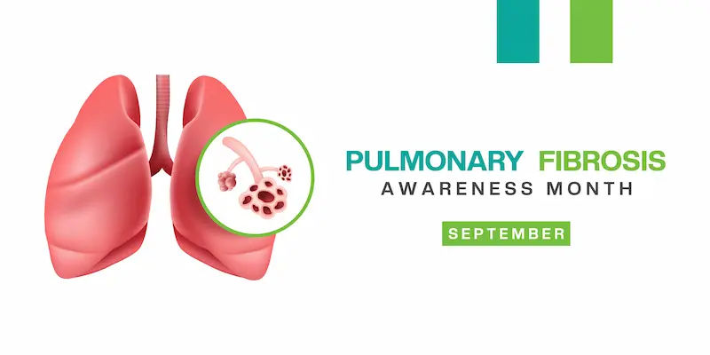
Guide to Lung Fibrosis
Learn about lung fibrosis (pulmonary fibrosis)—its symptoms, causes, diagnosis, and treatment options. Explore care strategies, pulmonary rehab, and lifestyle management tips to live well with fibrosis.


Introduction
Lung fibrosis—often called pulmonary fibrosis—means scarring inside the lungs that makes it harder to breathe and deliver oxygen to the body. While “fibrosis” sounds technical, many people first notice everyday changes: shortness of breath on stairs, a cough that won’t go away, or fatigue they can’t shake. This guide translates the science into practical steps you can use. We’ll explain what lung fibrosis is, how it’s diagnosed, and the treatments that can slow progression. You’ll learn the difference between idiopathic pulmonary fibrosis (IPF) and other interstitial lung diseases, what tests to expect, and how pulmonary rehabilitation, oxygen therapy, and lifestyle changes can help. We also cover advanced care options like lung transplant, and how to plan for travel, work, and daily life. If symptoms persist beyond two weeks or worsen, consult a doctor online with Apollo 24|7 for further evaluation. Whether you’re newly diagnosed or supporting a loved one, this comprehensive, evidence-informed guide puts you in control of your next steps.
What Is Lung Fibrosis?
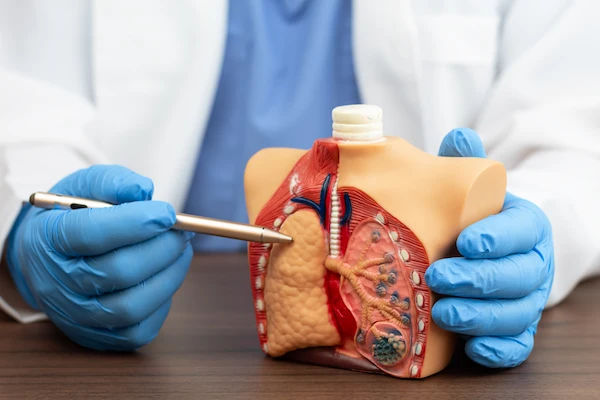
Lung fibrosis is the formation of excess scar tissue in the lungs. Over time, this scarring stiffens the delicate air sacs (alveoli), reducing the lungs’ ability to transfer oxygen into the bloodstream. The result is breathlessness, cough, and exercise intolerance. While some people use “pulmonary fibrosis” and “interstitial lung disease” (ILD) interchangeably, they aren’t identical. ILD is a broader category that includes dozens of conditions affecting the interstitium (the lung’s supporting framework). Many ILDs involve inflammation that can be reversible early on; others, particularly those described as “fibrosing,” lead to permanent scarring.
Pulmonary fibrosis may be “idiopathic” (IPF) when no clear cause is found, or “secondary” to exposures (like silica or mold), autoimmune diseases (such as rheumatoid arthritis), certain medications, or prior infections. Idiopathic pulmonary fibrosis is a distinct clinical entity with a characteristic pattern on high-resolution CT (HRCT) called usual interstitial pneumonia (UIP) and specific management strategies.
What changes inside the lung? In fibrosis, normal tissue is replaced by collagen-rich scar that thickens the walls of the air sacs. This increases stiffness (reduced compliance) and decreases the lung’s diffusion capacity (DLCO). The body compensates by increasing breathing rate, but activities like walking or climbing may still trigger oxygen desaturation. Understanding this physiology explains why treatments aim to slow scarring, maintain conditioning, and ensure adequate oxygen delivery.
Common Symptoms and When to Seek Help
The most common symptoms include:
- Shortness of breath, first with exertion, later at rest
- A persistent dry cough, often worse at night or with talking
- Fatigue, reduced exercise tolerance
- Crackling “Velcro-like” sounds heard by clinicians on auscultation
- Unintentional weight loss or chest discomfort in some cases
- Clubbing (widening) of the fingertips in advanced disease
Red flags needing prompt evaluation:
- Rapidly worsening breathlessness
- Chest pain, fainting, or new palpitations
- Blue lips or fingertips (cyanosis)
- High fever with cough and sputum suggesting infection
If you notice persistent shortness of breath or cough beyond two weeks—especially if it limits your daily activities—consult a doctor online with Apollo 24|7 for further evaluation. Prepare for your visit by noting when symptoms started, what makes them better/worse, any environmental exposures (dust, birds, molds), medication history (including prior chemotherapy or amiodarone), and family history of lung disease.
Causes and Risk Factors You Should Know
Lung fibrosis arises from varied causes:
- Genetics and family history: Some families have a higher risk due to variants in genes involved in telomere
maintenance and mucin production. If multiple first-degree relatives have pulmonary fibrosis, mention this to your
clinician. - Environmental/occupational exposures: Long-term exposure to silica, asbestos, metal dusts, wood dust, agricultural dust, birds (avian proteins), and molds can trigger fibrosing ILDs such as hypersensitivity pneumonitis.
- Autoimmune diseases: Rheumatoid arthritis, systemic sclerosis (scleroderma), and mixed connective tissue disease can cause ILD. Clues include joint pain, Raynaud’s phenomenon, skin thickening, or dry eyes/mouth.
- Medications and radiation: Some chemotherapy agents, nitrofurantoin, and amiodarone have been linked to lung scarring.
- Infections and post-viral changes: Severe pneumonia and, in some, post-COVID changes can lead to persistent interstitial abnormalities. Many post-viral changes improve over months, but a subset may develop residual fibrosis—follow-up imaging/PFTs guide care.
Lifestyle factors and comorbidities:
- Smoking increases risk and can worsen outcomes.
- Reflux disease (GERD) is common in IPF; managing reflux may reduce cough and aspiration risk, though routine antacid therapy solely to slow fibrosis has mixed evidence.
How Lung Fibrosis Is Diagnosed
Diagnosis blends clinical history, specialized imaging, lung function testing, and sometimes tissue sampling:
- High-resolution CT (HRCT): The key imaging test. A “UIP pattern”—basal/subpleural predominant reticulation, traction bronchiectasis, and honeycombing—supports IPF in the right clinical context. Other patterns suggest alternative causes (e.g., NSIP pattern in autoimmune ILD).
- Pulmonary function tests (PFTs): Measure forced vital capacity (FVC) and diffusion capacity (DLCO). ILD often shows a restrictive pattern (reduced FVC) and reduced DLCO. A 6-minute walk test assesses oxygen desaturation and functional capacity.
- Blood tests: Check for autoimmune markers (ANA, RF, anti-CCP, myositis panel) and, in select cases, hypersensitivity panels. Apollo 24|7 offers convenient home collection for tests like ANA and rheumatoid factor, helping you avoid extra trips if breathing is difficult.
- Biopsy: Bronchoscopy with transbronchial biopsies or cryobiopsy may be considered; surgical lung biopsy is less common today when HRCT and clinical findings are sufficiently characteristic.
Multidisciplinary discussion (MDD): A team of pulmonologists, radiologists, and pathologists review the data together to refine diagnosis and guide management. This “team read” reduces misdiagnosis and avoids unnecessary biopsies.
Staging, Progression, and Prognosis
Unlike cancer, lung fibrosis isn’t staged numerically the same way, but severity and progression are gauged by:
- Lung function: FVC and DLCO trends over time
- Oxygen needs at rest and with exertion
- HRCT extent of fibrosis
- Symptoms and exercise tolerance (6-minute walk distance)
Progressive pulmonary fibrosis (PPF): Many non-IPF ILDs can behave like IPF, with ongoing scarring despite treatment. PPF is defined by worsening symptoms, declining lung function, and/or increasing fibrosis on imaging over a set period (often 12 months). Identifying PPF matters because antifibrotic therapy (like nintedanib) can slow decline in various progressive fibrosing ILDs.
Acute exacerbations: Sudden worsening in breathlessness with new diffuse lung opacities on CT may represent an “acute exacerbation” and requires urgent care. Survival after exacerbations is variable; preventing infections, staying up to date on vaccinations, and early care for worsening symptoms may help reduce risks.
Monitoring frequency: Many clinics check PFTs every 3–6 months and HRCT annually or when symptoms change.
Home pulse oximeters and symptom diaries can detect early trends.
Treatment Options: From Medicines to Oxygen
Antifibrotic medications:
- Pirfenidone: In the ASCEND trial, pirfenidone reduced the proportion of patients with significant FVC decline or death
over 52 weeks compared with placebo. - Nintedanib: In the INPULSIS trials, nintedanib slowed annual FVC decline in IPF patients. The INBUILD trial later showed nintedanib slowed lung function decline in diverse progressive fibrosing ILDs, not just IPF.
Side effects vary (pirfenidone: photosensitivity, GI upset; nintedanib: diarrhea, liver enzyme elevations). Your clinician will monitor labs and help manage tolerability.
Treating underlying causes:
- Autoimmune ILD may improve with immunosuppressive therapy when inflammation is predominant. When scarring dominates or progression continues, antifibrotics may be considered.
- Reflux management: Positioning, diet changes, and individualized medical therapy may reduce reflux-related microaspiration that can worsen cough.
Oxygen therapy and vaccinations:
- Oxygen supplementation improves exercise tolerance and quality of life when oxygen levels drop with activity or at rest. Your team will titrate flow to keep saturation typically above 88–90% during exertion.
- Vaccinations (influenza, pneumococcal, COVID-19 per local guidance) reduce infection risk—an important step in preventing exacerbations.
Clinical trials and emerging treatments:
- Ongoing research targets multiple fibrosis pathways. If options feel limited, ask about clinical trial eligibility at any stage.
If your condition does not improve after trying prescribed treatments, book a physical visit to a doctor with Apollo 24|7 for a thorough reassessment.
Consult Top Specialists Here
Pulmonary Rehabilitation and Everyday Self-Care
Pulmonary rehabilitation (PR) is a supervised program combining exercise, education, and support. In ILD, PR improves walking distance, breathlessness, and quality of life. A typical PR plan includes:
- Aerobic training: Interval or continuous walking/cycling tailored to your oxygen needs.
- Strength training: Especially lower limbs, to reduce exertional breathlessness.
- Breathing techniques: Pursed-lip breathing and paced breathing help control dyspnea.
- Energy conservation: Practical tips like sit-to-stand strategies, shower stools, and planning chores during “good energy windows.”
Home strategies:
- Activity pacing: Break tasks into smaller chunks, rest before you feel exhausted, and use “breath breaks.”
- Nutrition and hydration: Adequate protein preserves muscle; smaller, frequent meals reduce post-meal breathlessness.
- Sleep quality: Elevate the head of your bed slightly if reflux is an issue; treat sleep apnea if present.
- Air quality: Avoid smoke, occupational dust, and indoor molds; consider HEPA filtration in frequently used rooms.
Travel and altitude:
- Air travel cabins are pressurized to the equivalent of 6,000–8,000 feet; if you desaturate with walking at home, discuss in-flight oxygen. Many airlines require medical clearance forms.
- High-altitude destinations may worsen hypoxemia; plan oxygen and rest breaks and consider alternative venues when feasible.
Managing Complications and Flare-ups
Common complications:
- Infections: Viral or bacterial infections can tip stable disease into a flare. Early recognition (fever, change in cough/sputum, increased breathlessness) and prompt treatment matter. Hand hygiene, vaccines, and avoidance of sickcontacts reduce risk.
- Pulmonary hypertension: Long-standing low oxygen can raise pressure in lung arteries, worsening breathlessness.
- Echocardiography screens for this; oxygen and targeted therapies may help select patients.
- Sleep apnea: Common and treatable; CPAP can improve daytime energy and cognition.
- Persistent cough: Strategies include treating reflux, humidification, cough suppression techniques, and prescribed medications as needed.
When to get urgent care:
- Sudden, significant worsening in breathlessness
- Chest pain, fainting, or new leg swelling (possible blood clots)
- Blue lips/fingertips or confusion (low oxygen)
If any of these occur, seek urgent medical care immediately. If you’re unsure whether a change is serious, consult a doctor online with Apollo 24|7 for rapid guidance and triage.
Advanced Care Planning: Transplant and Palliative Support
Lung transplant:
- Who and when: People with progressive disease, rising oxygen needs, or steeply declining FVC/DLCO should discuss early referral—even if they still feel “okay.” Evaluation takes time, and listing before a crisis improves outcomes.
- What to expect: Pre-transplant assessments, optimisation (nutrition, rehab), and post-transplant life with immunosuppression and frequent follow-up.
- Outcomes: Survival after transplant has improved over time; for appropriate candidates, it can restore independence and exercise capacity.
Palliative care:
- Symptom-focused care (breathlessness, cough, anxiety, insomnia) is beneficial at any stage and can be integrated alongside disease-modifying therapy.
- Advance directives: Document your preferences and share them with family and your care team.
Living Well with Lung Fibrosis
Building your support network:
- Join local or online groups through reputable organizations (e.g., disease foundations). Peer tips can be practical and
morale-boosting.
Tools and tracking:
- Apps or simple spreadsheets to log symptoms, step counts, oximetry, and inhaler/med schedules reveal patterns you can act on.
Work and financial planning:
- Ask HR about accommodations (flexible hours, remote days, reduced physical demands). Investigate disability benefits early if needed to avoid gaps in coverage.
Conclusion
Lung fibrosis can feel overwhelming at first, but knowledge and a supportive care plan make a real difference. Start with a clear diagnosis—often via HRCT, lung function tests, and a multidisciplinary review—so your team can tailor therapy. If you have idiopathic pulmonary fibrosis or progressive pulmonary fibrosis, antifibrotic medications may slow decline, while pulmonary rehabilitation, oxygen optimisation, and infection prevention can help you stay active and independent. Keep a simple tracking routine for symptoms and oxygen levels, practice energy-conserving habits, and build a network that includes family, peers, and clinicians. If your symptoms persist or worsen, consult a doctor online with Apollo 24|7 for timely assessment; Apollo 24|7 also offers convenient home collection for common lab tests related to autoimmune screening. Above all, check in regularly about your goals—from travel to work to family time—so your care plan stays aligned with what matters most. You’re not alone, and with proactive steps, you can live fully while navigating lung fibrosis.
Consult Top Specialists Here
Consult Top Specialists Here

Dr. Aakanksha Chawla
Pulmonology Respiratory Medicine Specialist
9 Years • MD (Pulmonary Medicine), IDCCM, IFCCM (Indian Fellowship in Critical Care Medicine)
Delhi
Apollo Hospitals Indraprastha, Delhi
(275+ Patients)

Dr. Sumara Maqbool
Pulmonology Respiratory Medicine Specialist
12 Years • MBBS, DNB Respiratory, critical care and sleep medicine, DrNB superspeciality Critical care, IDCCM, IFCCM, EDIC
Delhi
Apollo Hospitals Indraprastha, Delhi
(25+ Patients)

Dr Vishwa Vijeth K.
Pulmonology Respiratory Medicine Specialist
8 Years • MBBS, MD ( Respiratory Medicine)
Bangalore
Apollo Clinic Bellandur, Bangalore

Santoshkumar P Hammigi
Pulmonology Respiratory Medicine Specialist
4 Years • MBBS,MD, (Respiratory Medicine)
Bengaluru
Apollo Medical Center, Marathahalli, Bengaluru
(25+ Patients)

Dr. Chaithanya R
Internal Medicine Specialist Diabetologist
16 Years • MBBS, MD Internal Medicine, Fellowship in Diabetes(UK), CCEBDM(PHFI)
Bangalore
Apollo Clinic Bellandur, Bangalore
(100+ Patients)
Consult Top Specialists Here
Dr. Aakanksha Chawla
Pulmonology Respiratory Medicine Specialist
9 Years • MD (Pulmonary Medicine), IDCCM, IFCCM (Indian Fellowship in Critical Care Medicine)
Delhi
Apollo Hospitals Indraprastha, Delhi
(275+ Patients)
Dr. Sumara Maqbool
Pulmonology Respiratory Medicine Specialist
12 Years • MBBS, DNB Respiratory, critical care and sleep medicine, DrNB superspeciality Critical care, IDCCM, IFCCM, EDIC
Delhi
Apollo Hospitals Indraprastha, Delhi
(25+ Patients)
Dr Vishwa Vijeth K.
Pulmonology Respiratory Medicine Specialist
8 Years • MBBS, MD ( Respiratory Medicine)
Bangalore
Apollo Clinic Bellandur, Bangalore
Santoshkumar P Hammigi
Pulmonology Respiratory Medicine Specialist
4 Years • MBBS,MD, (Respiratory Medicine)
Bengaluru
Apollo Medical Center, Marathahalli, Bengaluru
(25+ Patients)
Dr. Chaithanya R
Internal Medicine Specialist Diabetologist
16 Years • MBBS, MD Internal Medicine, Fellowship in Diabetes(UK), CCEBDM(PHFI)
Bangalore
Apollo Clinic Bellandur, Bangalore
(100+ Patients)
More articles from Fibrosis Of Lung
Frequently Asked Questions
1) Is lung fibrosis the same as interstitial lung disease?
Not exactly. Interstitial lung disease (ILD) is a group of conditions; many involve inflammation and/or scarring. Lung fibrosis refers to the scarring part. Idiopathic pulmonary fibrosis is one specific ILD that is primarily fibrosing.
2) What are the first signs of lung fibrosis I should watch for?
Early signs include shortness of breath on exertion and a persistent dry cough. If these symptoms persist beyond two weeks or limit daily activities, consult a doctor online with Apollo 24|7 for evaluation.
3) Can lung fibrosis be cured?
There’s no cure for most forms, but treatments can slow progression and improve quality of life. Antifibrotic therapy (pirfenidone or nintedanib) may reduce lung function decline in idiopathic pulmonary fibrosis and some progressive pulmonary fibrosis cases.
4) What tests diagnose lung fibrosis?
High-resolution CT (HRCT), pulmonary function tests (FVC and DLCO), a 6-minute walk test, and blood tests for autoimmune causes are common. Biopsy is sometimes needed. Apollo 24|7 offers home collection for selected blood tests to support your diagnostic workup.
5) When should I consider a lung transplant for pulmonary fibrosis?
Discuss early referral if your lung function declines, oxygen needs increase, or symptoms progress despite treatment. Evaluation takes time; earlier listing often leads to better outcomes.