
Guide to Introduction On Sialendoscopy
Discover Sialendoscopy with our introductory guide. Learn what it is, how it works, its benefits, and what to expect during the procedure for salivary gland issues.

Introduction
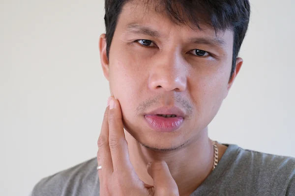
That sudden, sharp pain and swelling in your cheek or under your jaw while eating can be alarming. Often, this frustrating symptom points to a blocked salivary gland, a common issue traditionally treated with invasive surgery that risked scarring and nerve damage. But what if there was a way to fix the problem from the inside out, preserving your gland and leaving no visible marks? This is where sialendoscopy comes in. This article serves as your comprehensive introduction to sialendoscopy, a groundbreaking minimally invasive procedure that has revolutionized the treatment of salivary gland disorders. We will guide you through what it is, how it works, who it's for, and what you can expect, empowering you with the knowledge to discuss modern treatment options with your doctor confidently.
What Are Salivary Gland Obstructions and Why Do They Hurt?
To understand why sialendoscopy is so effective, we must first understand the problem it solves. Your salivary glands are essential for oral health, producing saliva that moistens food, aids digestion, and protects your teeth from decay.
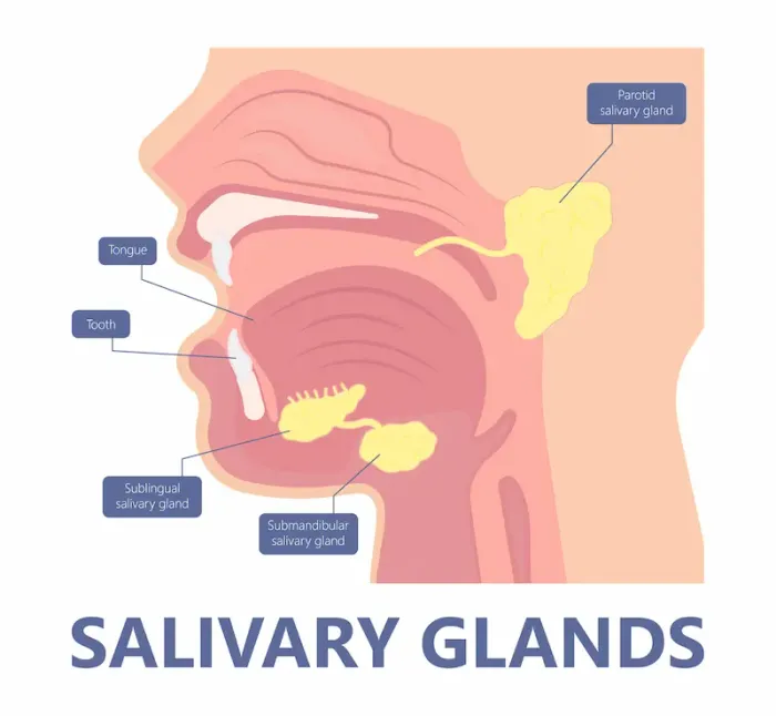
Meet Your Salivary Glands: The Unsung Heroes of Digestion
You have three pairs of major salivary glands:
• Parotid Glands: Located in front of your ears, these are the largest.
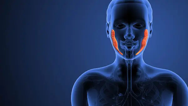
• Submandibular Glands: Situated under your jawbone, these produce most of your saliva and are the most common site for stones.
• Sublingual Glands: Found under the tongue.
Tiny ducts act as pipes, carrying the saliva from these glands into your mouth.
Common Culprits: Stones, Strictures, and Blockages
The most common cause of obstruction is a salivary stone, or sialolith. These are calcified masses that form from minerals in saliva, much like kidney stones. When you eat and your salivary glands are stimulated, saliva production increases. If a stone blocks the duct, the saliva has nowhere to go, leading to a painful backup and swelling known as mealtime syndrome. Other blockages can include strictures (narrowing of the duct) or mucous plugs.
Consult Top Specialists
What is Sialendoscopy? The "Keyhole" Revolution for Your Glands
Sialendoscopy is a minimally invasive endoscopic technique that allows an ENT surgeon to diagnose and treat salivary gland obstructions directly within the ductal system. Think of it as a tiny, high-tech camera journey into the salivary ducts.
The Sialendoscope: A Marvel of Miniaturized Technology
The core instrument is the sialendoscope itself—an ultra-thin, flexible or semi-rigid fiber-optic scope. It’s often less than 1.2 mm in diameter (about the size of a thick piece of spaghetti). This scope contains a miniature camera, a light source, and an irrigation channel. It is gently inserted into the natural opening of the salivary duct in the mouth, allowing the surgeon to navigate the intricate ductal network in real-time on a monitor. This direct visualization is what sets it apart, transforming guesswork into precise intervention.
Are You a Candidate? Signs and Symptoms That Point to Sialendoscopy
You might be a candidate for sialendoscopy if you experience recurrent episodes of:
1. Painful swelling in the cheek or under the jaw, especially when eating.
2. Tenderness and redness in the area.
3. Recurrent salivary gland infections (sialadenitis).
4. A bad taste in the mouth or pus drainage.
If these symptoms persist beyond two weeks, it's crucial to consult a doctor for an accurate diagnosis. If your condition does not improve after trying conservative methods like hydration and massage, book a physical visit to an ENT specialist with Apollo24|7 for further evaluation.
The Sialendoscopy Procedure: What to Expect, Step by Step
Understanding the process can alleviate much of the anxiety associated with any medical procedure.
Step 1: Diagnosis and Pre-Procedure Planning
Your doctor will first confirm the obstruction using imaging like an ultrasound or a CT scan. This helps locate the stone or identify the structure, planning the best approach for the sialendoscopy procedure.
Step 2: The Day of the Procedure – Anesthesia and Setup
Sialendoscopy is typically performed as an outpatient procedure under local or general anesthesia, depending on the complexity. The mouth is kept open with a retractor, and the surgeon will locate the natural duct opening, often dilating it slightly for scope insertion.
Step 3: The Guided Journey Inside the Salivary Duct
The sialendoscope is carefully advanced into the duct. Saline solution is flushed through the irrigation channel to expand the duct, providing a clear view. The surgeon navigates through the ductal system, looking for the source of the blockage.
Tools of the Trade: Baskets, Balloons, and Lasers
Once the obstruction is found, miniature tools are passed through a working channel in the scope:
• Micro Baskets: Used to ensnare and retrieve stones.
• Balloons: Inflated to dilate strictures and widen the duct.
• Micro Lasers: Can fragment larger stones that are too big to remove whole.
This minimally invasive salivary stone removal is the hallmark of the procedure.
Step 4: Immediate Aftermath and Recovery Room
After the blockage is cleared, the scope is removed. The procedure typically lasts between 30 to 90 minutes. Patients are monitored for a short period before being discharged, usually on the same day.
Sialendoscopy vs. Traditional Surgery: A Clear-Cut Advantage
For decades, the only solution for persistent stones was open gland surgery (e.g., submandibulectomy or parotidectomy). This involved an external incision in the neck or face, removal of the stone, and sometimes the entire gland. This approach carried risks of:
1. Visible scarring.
2. Damage to facial nerves, potentially leading to permanent facial weakness.
3. Longer recovery times.
4. Dry mouth (xerostomia) if the gland was removed.
Preserving Your Gland, Avoiding Scars: The Core Benefits
Sialendoscopy offers a paradigm shift with distinct advantages:
• Gland Preservation: The primary goal is to save the gland and its function.
• No External Scars: All access is through the natural duct opening in the mouth.
• Reduced Risk: Significantly lower risk of nerve damage.
• Faster Recovery: Patients often return to normal activities within a day or two.
• High Accuracy: Direct visualization ensures precise treatment.
Potential Risks and Realistic Success Rates
No procedure is without risk. Potential but rare complications of sialendoscopy include duct perforation, infection, or instrument breakage. However, in experienced hands, the procedure is exceptionally safe. Success rates are high, often exceeding 80-95% for stone removal and stricture dilation, making it the gold-standard first-line treatment for obstructive salivary gland diseases.
Life After Sialendoscopy: Recovery and Long-Term Care
The recovery time after sialendoscopy is typically swift. You may experience mild discomfort or swelling, managed with over-the-counter pain relievers. Your doctor will advise you to maintain excellent oral hygiene, stay hydrated, and may recommend sucking on sour candies to stimulate saliva flow and keep the ducts open. Most patients experience immediate relief from their symptoms.
Consult Top Specialists
Conclusion
Chronic salivary gland pain can significantly impact your quality of life, turning every meal into a source of anxiety. The advent of sialendoscopy has transformed this landscape, offering a precise, effective, and gentle solution. By addressing the root cause of the obstruction without the downsides of traditional surgery, it represents a significant leap forward in patient care. If you recognize the symptoms discussed in this introduction to sialendoscopy, don't hesitate to seek expert advice. Modern medicine has a powerful tool to help you regain comfort and health. Consult a doctor online with Apollo24|7 to discuss your symptoms and explore whether a referral to an ENT specialist is the right next step for you.
Consult Top Specialists

Dr. Impana G N
Physician/ Internal Medicine/ Covid Consult
11 Years • MBBS,DNB FAMILY MEDICINE, MNAMS ,CCEBDM
Mysuru
Apollo BGS Hospital Adichuchanagiri Road, Mysuru

Dr. M. Krishna Bharath Reddy
General Physician/ Internal Medicine Specialist
10 Years • MD General Medicine, MRCP (London) and DM( Critical Care)
Chennai
Apollo One Chennai, Chennai

Dr. Moumita Roy
General Physician/ Internal Medicine Specialist
8 Years • MBBS , MD (Anesthesiology)
Kolkata
VDC Clinic, Kolkata

Dr. Soumen Paul
General Practitioner
24 Years • MBBS
Kolkata
MCR SUPER SPECIALITY POLY CLINIC & PATHOLOGY, Kolkata
(25+ Patients)

Dr. Mijanur Rahaman Mondal
General Practitioner
3 Years • MBBS
Kolkata
Dr Utsa Basu Clinic, Kolkata
(25+ Patients)
Consult Top Specialists
Dr. Impana G N
Physician/ Internal Medicine/ Covid Consult
11 Years • MBBS,DNB FAMILY MEDICINE, MNAMS ,CCEBDM
Mysuru
Apollo BGS Hospital Adichuchanagiri Road, Mysuru
Dr. M. Krishna Bharath Reddy
General Physician/ Internal Medicine Specialist
10 Years • MD General Medicine, MRCP (London) and DM( Critical Care)
Chennai
Apollo One Chennai, Chennai
Dr. Moumita Roy
General Physician/ Internal Medicine Specialist
8 Years • MBBS , MD (Anesthesiology)
Kolkata
VDC Clinic, Kolkata
Dr. Soumen Paul
General Practitioner
24 Years • MBBS
Kolkata
MCR SUPER SPECIALITY POLY CLINIC & PATHOLOGY, Kolkata
(25+ Patients)
Dr. Mijanur Rahaman Mondal
General Practitioner
3 Years • MBBS
Kolkata
Dr Utsa Basu Clinic, Kolkata
(25+ Patients)
More articles from Salivary gland disorders

Frequently Asked Questions
1. Is sialendoscopy painful?
The procedure is performed under anesthesia, so you will not feel pain during it. Afterwards, most patients experience only mild discomfort, similar to a toothache, which is easily managed with medication.
2. What is the typical cost of a sialendoscopy procedure?
The cost varies widely based on geographic location, hospital facility, and the complexity of the case. It's best to consult directly with an ENT specialist and your insurance provider for a precise estimate.
3. Can all salivary stones be removed with sialendoscopy?
While highly effective, very large or oddly shaped stones embedded deep within the gland might require a combined approach, using sialendoscopy alongside other minor surgical techniques. However, it is successful in the vast majority of cases.
4. Are there any dietary restrictions after sialendoscopy?
You may be advised to stick to soft foods for a day or two. The most important dietary advice is to stay well-hydrated and avoid foods that overly stimulate saliva production immediately after the procedure, as this can cause discomfort.
5. How do I find the best doctor for sialendoscopy?
Look for an Ear, Nose, and Throat (ENT) specialist, specifically an otolaryngologist, who has fellowship training or significant experience in salivary gland disorders and interventional sialendoscopy. Academic medical centers are often at the forefront of this technology.