Guide to Interventional Neurology Neurosciences/brain Attack Or Stroke Emergency
Learn the BE-FAST stroke warning signs and why every minute counts during a brain attack. This guide covers interventional neurology options like IV thrombolysis and mechanical thrombectomy that rapidly restore blood flow to the brain, saving function and improving recovery.


Introduction
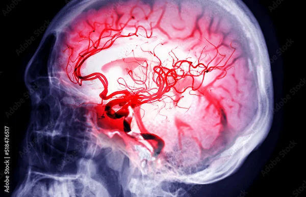
A stroke is often called a “brain attack” for good reason—when blood flow to the brain stops, brain cells start dying within minutes.That’s why interventional neurology—advanced, minimally invasive treatments delivered through blood vessels—is one of the most important breakthroughs in modern neurosciences/brain care. Today, specialists can dissolve clots with medicines and even remove them with tiny devices inside an artery, restoring blood flow and saving brain function.
This guide walks you through stroke warning signs, what to do in an emergency, and exactly how interventional neurology works in the first hours after a brain attack. You’ll learn about clot-busting drugs, mechanical thrombectomy, advanced imaging, and what happens in the hospital during a “stroke code.” We’ll also cover recovery, prevention, and how technologies like telestroke and AI speed up lifesaving care. Whether you’re reading for yourself or a loved one, the goal is simple: recognise stroke fast, act faster, and understand the interventional neurology options that protect the brain.
Brain Attack: Why Every Minute Matters
Recognising stroke symptoms immediately and calling emergency services is the single most important action to save
brain tissue.
Stroke occurs when the brain’s blood supply is blocked (ischaemic stroke)or a blood vessel bursts (haemorrhagic stroke).
About 85% are ischaemic; in these, a clot stops blood flow, starving brain tissue of oxygen and nutrients. The rest are
haemorrhagic, often from long-standing high blood pressure or ruptured aneurysms. Either way, “time is brain.” Faster
treatment means more brain saved and better recovery chances.
The BE-FAST Checklist
Use the BE-FAST checklist to recognise stroke symptoms quickly:
- B: Balance—sudden dizziness, trouble walking
- E: Eyes—sudden vision loss or double vision
- F: Face—one side droops
- A: Arms—one arm drifts or feels weak
- S: Speech—slurred speech or confusion
- T: Time—call emergency services immediately
Do not self-drive to the hospital; emergency medical services can pre-alert a stroke centre, ensuring CT scans and stroke
teams are ready on arrival. Evidence shows that early treatment with clot-busting drugs within 4.5 hours and
mechanical thrombectomy for large artery blockages can dramatically improve outcomes. The earlier treatment starts,
the greater the benefit. If someone shows BE-FAST signs even if they improve, treat it as a brain attack and activate
emergency response right away.
What Is Interventional Neurology?
Interventional neurology uses minimally invasive catheter-based techniques to treat blood vessel disorders from inside
the arteries.
Interventional neurology (also called neurointerventional surgery or endovascular neurosurgery) uses catheters and
imaging to treat brain and spine blood vessel disorders from inside the artery, without open surgery. For acute
ischaemic stroke, the goal is to reopen blocked arteries rapidly. In many hospitals, a “stroke code” activates a team that
includes emergency physicians, neurologists, neurointerventionalists, anaesthesiologists, nurses, and radiology
technologists.
Consult Top Neurologists for Personalised Advice
The Stroke Code Flow
Here’s how a stroke code flows:
- Pre-notification and Triage
- Pre-notification: EMS alerts the hospital of suspected stroke and the last-known-well time.
- Rapid triage: On arrival, a focused exam and BE-FAST screen guide immediate non-contrast CT to rule out bleeding.
- Imaging and Decision
- Parallel processing: If ischaemic stroke is suspected, labs, vital signs, and IV access are obtained while the patient heads
to imaging; the team reviews contraindications for thrombolysis. - Vascular imaging: CT angiography identifies large vessel occlusions (LVO). If LVO is present and within criteria, the
neurointerventional team is paged for mechanical thrombectomy. - Decision and consent: The stroke team communicates risks, benefits, and alternatives to the patient/family quickly and
clearly.
Interventional neurology also treats haemorrhagic strokes (e.g., aneurysm coiling) and reduces future risk (e.g., carotid
stenting). But in a brain attack, the central mission is to restore blood flow quickly and safely—a core focus across
neurosciences/brain emergency care.
Getting to the Right Treatment Fast
Choosing the correct hospital pathway and shaving minutes off treatment times is critical for good patient outcomes.
Calling an ambulance is not just about transportation. Paramedics can check glucose (to rule out a stroke mimic),
monitor airway and blood pressure, and triage to the nearest stroke-ready hospital. Many regions have protocols to
route suspected LVO cases directly to a comprehensive stroke centre, where mechanical thrombectomy is available.
This “right place, first time” strategy shortens the time to “door-to-groin puncture” (start of the procedure) for
endovascular therapy.
Two Speed Metrics Matter
- Door-to-needle time: time from hospital arrival to IV thrombolysis. The target is typically within 60 minutes; faster is
better. - Door-to-groin puncture: time to start thrombectomy. High-performing centres achieve 60–90 minutes, often faster with
pre-alerts.
Telestroke programmes allow neurologists to evaluate patients by video in smaller hospitals, guiding decisions on
thrombolysis and transfer. Some cities deploy mobile stroke units—ambulances with CT scanners—to start treatment in
the field. These innovations shrink time to treatment and improve outcomes. A unique tip: save the exact “last-known-
well” time in your phone and share medications (especially blood thinners) with EMS.
Intravenous Clot-Busting: Alteplase and Tenecteplase
Intravenous thrombolysis uses medication delivered into a vein to dissolve the clot, often serving as the first-line or
"bridging" therapy.
For many ischaemic strokes, intravenous thrombolysis is the first-line therapy. Alteplase (tPA) is approved within 4.5
hours of symptom onset in eligible patients; earlier is better. Eligibility excludes active bleeding, recent major surgery,
very high blood pressure unresponsive to medication, or certain lab abnormalities. The main risk is bleeding, including
intracranial haemorrhage, but careful selection makes the benefits outweigh the risks for many patients.
Tenecteplase (TNK) as an Alternative
Tenecteplase (TNK) is a related clot-busting medicine given as a single IV bolus. Several studies suggest TNK can be at
least as effective as alteplase for early reperfusion, especially when thrombectomy is planned, with logistical advantages
because it’s a one-time push rather than an hour-long infusion. Current guidelines in many regions allow TNK as a
reasonable alternative to alteplase in specific scenarios. The choice depends on hospital protocols, timing, clot location,
and imaging.
Bridging Therapy Example
Real-world example: A patient with sudden right-sided weakness arrives 70 minutes after last known well. CT shows no
bleed; the team administers TNK within 20 minutes of arrival and moves directly to angiography for possible
thrombectomy. This “bridging therapy” can dissolve smaller clots and improve microcirculation while the
interventional team prepares. If symptoms persist beyond the initial treatment period or if complications arise, close
neurocritical care monitoring guides the next steps.
Mechanical Thrombectomy: Modern Endovascular Therapy
This procedure involves using catheter-based devices to physically extract large clots from major arteries in the brain.
Mechanical thrombectomy is a minimally invasive procedure that uses a catheter threaded through an artery (usually in
the groin or wrist) to remove a clot from a large brain artery. Devices include stent retrievers and aspiration catheters
that trap and extract the clot, restoring blood flow. This approach is highly effective for large vessel occlusions—
commonly the internal carotid artery or proximal middle cerebral artery
Eligibility and Time Window
Landmark trials demonstrated that, in selected patients treated within roughly 6 hours, thrombectomy markedly
improves functional outcomes compared with medical therapy alone. Later trials showed that with advanced imaging
to identify salvageable brain tissue (the “penumbra”), selected patients benefit even when treated 6–24 hours after last-
known-well. In practice, this means that even if you wake up with stroke symptoms, you might still be eligible for
thrombectomy after imaging.
Quality Metrics
A unique perspective: Ask your team about “first-pass effect”—opening the artery on the first attempt is associated with
better outcomes. High-volume centres track this metric along with time intervals and reperfusion grades (e.g., TICI
2b/3). These details reflect a culture of quality and can influence recovery.
Imaging the Brain in an Emergency
Rapid, multi-modal brain imaging is essential to confirm the diagnosis, rule out bleeding, and determine eligibility for
endovascular treatment.
Imaging drives stroke decisions. The first step is usually a non-contrast CT to rule out haemorrhage and look for early
signs of ischaemia. If no bleed is seen, CT angiography (CTA) maps the arteries to detect blockages. Many centres add
CT perfusion (CTP) to distinguish irreversibly damaged core tissue from salvageable penumbra. MRI/MRA may be
used when CT findings are equivocal or for posterior circulation strokes where early CT signs are subtle.
Imaging Answers Key Questions
- H3: Bleeding and Occlusion
- Is there bleeding? If yes, no thrombolysis; consider neurosurgical or endovascular treatment for aneurysm or AVM.
- Is there a large vessel occlusion? If yes, consider thrombectomy.
- H3: Penumbra and Mimics
- Is there mismatch between small core and large penumbra? If yes, consider late-window thrombectomy up to 24 hours.
- Are there stroke mimics (e.g., seizure, tumour)? Imaging plus labs and clinical assessment help avoid unnecessary
treatments.
A practical tip: The best imaging is the one you can get quickly. High-quality, fast CT/CTA/CTP at a stroke centre
often speeds decisions better than waiting for an MRI. In prehospital systems, seamless image sharing and AI-assisted
triage can alert interventional teams earlier, shaving precious minutes off door-to-groin times.
Special Scenarios in Stroke Care
Atypical presentations, especially in the posterior circulation, require high clinical suspicion and careful imaging to
ensure timely intervention.
Not all strokes present the same way. Posterior circulation strokes (brainstem or cerebellum) can start with dizziness,
nausea, double vision, or trouble walking. These can be missed if one looks only for face or arm weakness. The “BE-
FAST” addition (Balance and Eyes) is designed to catch these strokes earlier. CTA/MRA is very helpful here;
thrombectomy may be considered for basilar artery occlusion.
Stroke Mimics
Stroke mimics include hypoglycaemia, seizure with postictal paralysis (Todd’s paralysis), migraine with aura, functional
disorders, and infections. That’s why glucose is checked immediately and why imaging is essential. The aim is to give
life-saving therapy quickly to true strokes while avoiding unnecessary thrombolysis in mimics. If symptoms fluctuate or
rapidly improve (possible TIA), urgent evaluation is still needed; TIAs are powerful warnings that a larger stroke could
occur if risk factors aren’t addressed.
After the Emergency: Recovery, Rehab, and Risks
The immediate post-stroke phase focuses on monitoring the brain, preventing complications, and initiating early,
comprehensive rehabilitation.
Once blood flow is restored or stabilised, the focus shifts to limiting complications and optimising recovery. Patients
often go to a neuro intensive care unit or dedicated stroke unit, where teams watch blood pressure, oxygenation, blood
sugar, and temperature—each of which affects the injured brain. Early, tailored rehabilitation (physical, occupational,
speech therapy) begins as soon as it’s safe. Many patients see meaningful improvement in the first weeks to months.
Preventing Another Stroke Starts Immediately
- Atrial fibrillation: detection with continuous monitoring; anticoagulation when indicated lowers future stroke risk.
- Hypertension: aggressive control is vital.
- Cholesterol: high-intensity statins are often started.
- Diabetes: tight glucose control improves outcomes.
- Lifestyle: stop smoking, limit alcohol, maintain a healthy weight, and exercise as advised.
If you’re in India and need follow-up labs, Apollo 24|7 offers convenient home collection for tests like lipid profile and
kidney function. If your blood pressure or sugars remain uncontrolled after initial changes, consult a doctor online with
Apollo 24|7 for further optimisation and medication review.
Interventional Care Beyond Ischaemic Stroke
The catheter-based techniques of interventional neurology are also used to treat aneurysms, AVMs, and symptomatic
carotid disease.
Interventional neurology is not only for clots. For aneurysms (blood vessel bulges that can rupture), minimally invasive
options like coil embolisation or flow-diverting stents can prevent or stop bleeding. Arteriovenous malformations
(AVMs) sometimes require staged embolisations. For carotid artery disease, carotid stenting may be considered in select
patients as an alternative to surgery, particularly in those at high surgical risk. These procedures use the same catheter-
based pathways and imaging guidance as thrombectomy, with the advantage of smaller incisions, shorter hospital stays,
and quicker recoveries in many cases.
Multidisciplinary Decision-Making
A unique insight: Many decisions rely on multi-disciplinary “neurovascular boards” that include neuroradiology,
neurology, neurosurgery, and interventional experts—don’t hesitate to ask if your case will be reviewed by such a team.
Telestroke, AI, and Mobile Stroke Units
Technological advancements are expanding the reach of expert stroke care and significantly shortening crucial
treatment times.
New technology is changing stroke care. Telestroke connects smaller hospitals to stroke specialists by video for rapid
assessment and treatment decisions, reducing door-to-needle times. Some systems now use AI to analyse CT scans and
perfusion maps in minutes, flagging large vessel occlusions and notifying the interventional team while the patient is still
on the scanner table.
Mobile Stroke Units
Mobile stroke units (ambulances equipped with CT scanners and point-of-care labs) can start thrombolysis in the field,
cutting treatment times significantly and improving outcomes in certain regions. For rural and remote communities,
these tools bring tertiary neurosciences/brain expertise to the patient. The takeaway: even if you’re far from a
comprehensive stroke centre, calling EMS activates a network that can speed interventional neurology care to you.
Building Your Personal Stroke Plan
Proactive planning, including knowing personal risk factors and local resources, is vital for stroke prevention and
preparedness.
- Know your numbers: blood pressure, cholesterol. Keep them in your phone along with your medications.
- Learn BE-FAST and teach family and friends.
- Identify your nearest primary and comprehensive stroke centres.
- Create an emergency card: allergies, medications (especially blood thinners), key medical history, and emergency
contacts. - If you’ve had a TIA or minor stroke, ask about atrial fibrillation monitoring and whether you need a carotid ultrasound
or echocardiography. Apollo 24|7 can help coordinate follow-up visits and routine lab checks at home.
When to See a Doctor Online—and When Not To
Any suspicion of acute stroke requires immediate emergency service activation, while risk factor management can use
telemedicine.
Acute stroke is a 100% emergency—call your local emergency number immediately. Do not use telemedicine for a
suspected brain attack.
Telemedicine is Appropriate for:
- TIA evaluation after urgent in-person assessment
- Blood pressure, diabetes, and cholesterol management
- Reviewing imaging or reports after discharge
- Medication counselling and lifestyle planning
If you have recurrent brief neurologic episodes (possible TIAs), don’t wait. Seek urgent care the same day. For ongoing risk-factor control or if your condition does not improve after trying lifestyle changes, book a visit with a doctor through Apollo 24|7. For lab monitoring, Apollo 24|7 offers convenient home collection for lipid profile, $\text{HbA}_{1\text{c}}$, and thyroid/kidney tests that influence stroke risk.
Myths, Fears, and Realities
Dispelling common misconceptions about stroke helps ensure patients seek prompt, effective, and often life-saving
emergency care.
- Myth: “I’m too old for treatment.” Reality: Age alone is not an absolute barrier. Many older adults benefit from
thrombolysis and thrombectomy when eligible. - Myth: “If symptoms fade, it’s fine.” Reality: Symptoms that come and go can be TIAs; urgent evaluation prevents
major strokes. - Myth: “Thrombolysis always causes bleeding.” Reality: There’s risk, but selected patients have a net benefit, especially
when treated early. - Myth: “I’ll wait to see if it gets better.” Reality: Every minute equals millions of neurons—delays reduce benefit and increase disability.
Conclusion
Stroke care has transformed with the rise of interventional neurology. Today, many patients not only survive a brain attack but return to independent lives—if treatment starts fast. Recognising BE-FAST warning signs, calling emergency services immediately, and getting to a stroke-ready hospital are the keys that open the door to IV thrombolysis and mechanical thrombectomy. Imaging helps your team decide quickly and accurately, including whether you qualify for late-window therapies. After the emergency, rehabilitation and prevention—controlling blood pressure, cholesterol, diabetes, and atrial fibrillation—shape long-term outcomes.
Make a personal stroke plan: know your risks, your nearest stroke centres, and your medications. If you’re managing risk factors or need post-stroke follow-up, consult a doctor online with Apollo 24|7 and consider home collection for blood tests like lipid profiles. But in the face of sudden BE-FAST symptoms, there is only one right move: call emergency services now. Time saved is brain saved, and interventional neurology can make all the difference.
Consult Top Neurologists for Personalised Advice
Consult Top Neurologists for Personalised Advice

Dr Debnath Dwaipayan
Neurosurgeon
9 Years • MBBS, MS(Gen. Surgery), DrNB (Neurosurgery)
Delhi
Apollo Hospitals Indraprastha, Delhi

Dr. Sandeep Gurram
Neurologist
12 Years • MBBS, MD(Internal Medicine), DM(Neurology), PDF(Movement disorders) - NIMHANS
Secunderabad
Apollo Hospitals Secunderabad, Secunderabad

Dr. H Rahul
Neurologist
10 Years • MBBS, MD(Gen. Med.), DM(Neuro)
Secunderabad
Apollo Hospitals Secunderabad, Secunderabad
(100+ Patients)

Dr. Dipti Ranjan Tripathy
Neurologist
15 Years • MBBS, MD (GENERAL MEDICINE ),DM (NEUROLOGY)
Rourkela
Apollo Hospitals, Rourkela, Rourkela
(25+ Patients)

Dr. K Siva Rama Gandhi
Neurologist
11 Years • MBBS, DNB (Gen. Med.) DNB (Neurology) MBBS.,MD.,DNB
Kakinada
Apollo Hospitals Surya Rao Peta, Kakinada
(75+ Patients)
Consult Top Neurologists for Personalised Advice

Dr Debnath Dwaipayan
Neurosurgeon
9 Years • MBBS, MS(Gen. Surgery), DrNB (Neurosurgery)
Delhi
Apollo Hospitals Indraprastha, Delhi

Dr. Sandeep Gurram
Neurologist
12 Years • MBBS, MD(Internal Medicine), DM(Neurology), PDF(Movement disorders) - NIMHANS
Secunderabad
Apollo Hospitals Secunderabad, Secunderabad

Dr. H Rahul
Neurologist
10 Years • MBBS, MD(Gen. Med.), DM(Neuro)
Secunderabad
Apollo Hospitals Secunderabad, Secunderabad
(100+ Patients)

Dr. Dipti Ranjan Tripathy
Neurologist
15 Years • MBBS, MD (GENERAL MEDICINE ),DM (NEUROLOGY)
Rourkela
Apollo Hospitals, Rourkela, Rourkela
(25+ Patients)

Dr. K Siva Rama Gandhi
Neurologist
11 Years • MBBS, DNB (Gen. Med.) DNB (Neurology) MBBS.,MD.,DNB
Kakinada
Apollo Hospitals Surya Rao Peta, Kakinada
(75+ Patients)
More articles from Stroke
Frequently Asked Questions
1) What is the difference between a stroke and a brain attack?
They’re the same. “Brain attack” emphasises urgency, just as we say “heart attack.” Use the BE-FAST checklist and call emergency services immediately.75
2) How late is too late for mechanical thrombectomy?
Many patients benefit within 6 hours, and select patients identified by advanced imaging benefit up to 24 hours after last-known-well. Don’t delay—evaluation determines eligibility.
3) Is tenecteplase better than alteplase for stroke?
Both are effective clot busters. Tenecteplase (single bolus) is a reasonable alternative in select cases, especially when thrombectomy is planned.76 Protocols vary by centre.
4) Can I use telemedicine for stroke symptoms?
No. Stroke is an emergency—call your local emergency number.77 Use telemedicine later for TIA follow-up or risk-factor management. If your condition does not improve after initial measures, book a visit with Apollo 24|7.
5) What tests should I do after a stroke or TIA?
Your team may check cholesterol, $\text{HbA}_{1\text{c}}$, kidney function, thyroid, heart rhythm monitoring, carotid ultrasound, and echocardiography. Apollo 24|7 offers home collection for common labs that inform stroke prevention.


