A Patient's Guide to Fast and Accurate Diagnosis with Endoscopic Ultrasound (EUS)
Learn how Endoscopic Ultrasound (EUS) provides fast, accurate, and minimally invasive diagnosis for complex health conditions, combining imaging and biopsy in one procedure.

Written by Dr. J T Hema Pratima
Reviewed by Dr. Rohinipriyanka Pondugula MBBS
Last updated on 29th Sep, 2025

Introduction
Are you or a loved one facing a complex health issue where getting a clear, fast diagnosis feels like an endless wait? The uncertainty can be the hardest part. Modern medicine, however, has powerful tools to cut through that uncertainty. Endoscopic Ultrasound, or EUS, is one such advanced procedure that is revolutionising how doctors diagnose conditions deep within the body. This guide will demystify EUS, explaining in simple terms how it works, why it’s exceptionally effective for a speedy patient diagnosis, and what you can expect if your doctor recommends it. We'll explore how this "one-stop-shop" approach can simplify your diagnostic journey, providing clarity and paving the way for timely treatment.
What is Endoscopic Ultrasound (EUS)? Demystifying the Procedure
Imagine a doctor needing to examine your pancreas or the walls of your oesophagus. Standard scans like CT or MRI provide a good overview, but sometimes, they lack the fine detail needed for a definitive diagnosis. This is where Endoscopic Ultrasound (EUS) shines.
The Best of Both Worlds: Endoscopy Meets Ultrasound
An EUS procedure combines two powerful technologies:
- Endoscopy: A long, thin, flexible tube (an endoscope) is gently guided through your mouth or rectum to access your digestive tract.
- Ultrasound: A tiny ultrasound probe is attached to the tip of this endoscope. This probe emits sound waves that create detailed, high-resolution images of the surrounding organs and tissues.
Unlike a standard ultrasound on the skin, the EUS probe gets extremely close to the target area—like putting a microphone right next to a speaker instead of listening from the back of a hall. This proximity eliminates interference and produces incredibly clear images of structures that are otherwise difficult to see.
Why EUS is a Game-Changer for Internal Imaging
The key advantage of EUS is its ability to see beyond the surface. A standard endoscopy only shows the inner lining of the digestive tract. EUS, however, allows doctors to see through this lining to visualise the deeper layers of the gut wall, as well as adjacent organs like the pancreas, liver, gallbladder, and lymph nodes. This capability is fundamental to making a fast and accurate assessment of abnormalities.
How EUS Makes Your Diagnosis Faster and Simpler
EUS addresses diagnosis delays by streamlining the diagnostic process in two critical ways.
The "One-Stop-Shop" Advantage: Seeing and Sampling in One Procedure
This is perhaps the most significant benefit for a fast diagnosis. With many other tests, the process is sequential: a scan finds a suspicious area, and then a separate procedure (like a surgery or a percutaneous biopsy) is needed to collect a tissue sample (biopsy). This can take weeks.
Consult a Gastroenterologist for the best advice
With EUS, if the gastroenterologist sees a suspicious lesion, they can perform a EUS fine needle aspiration (FNA) or EUS fine needle biopsy (FNB) during the same procedure. A tiny needle is passed through the endoscope to collect cells or a tissue core from the target area. This "see-and-sample" approach condenses what used to be a multi-step, time-consuming process into a single session, dramatically speeding up the time to a definitive diagnosis.
Unparalleled Precision: Finding What Other Tests Miss
EUS is exceptionally sensitive. Studies have shown it to be superior to CT scans for detecting small tumours (especially in the pancreas) and for accurately staging cancers by assessing how deeply they have invaded a wall or if they have spread to nearby lymph nodes. For example, in pancreatic cancer, EUS can detect tumours as small as 2–3 mm, which might be invisible on other imaging modalities. This precision prevents the delays caused by inconclusive or false-negative results from less sensitive tests.
Common Conditions Where EUS Speeds Up Diagnosis
EUS is not used for every ailment, but for specific conditions, it is the gold standard.
Pancreatic and Bile Duct Issues: A Primary Application for EUS
The pancreas is located deep in the abdomen, behind the stomach, making it a challenging organ to image. EUS is the leading tool for:
- Evaluating Pancreatic Cysts and Tumours: It can distinguish between potentially cancerous and benign cysts with high accuracy.
- Investigating Unexplained Pancreatitis: It can identify tiny gallstones or other obstructions that other tests miss.
- Assessing Bile Duct Blockages: It helps determine if a blockage is due to a stone, a stricture, or a tumour.
Gastrointestinal Tumours: Staging Cancer with Remarkable Accuracy
For cancers of the oesophagus, stomach, and rectum, accurate staging (determining the extent of the cancer) is critical for planning treatment. EUS provides the most accurate assessment of how deep the cancer has grown into the organ wall and whether it has affected local lymph nodes. This information is vital for deciding if a tumour can be removed endoscopically or requires more extensive surgery.
Lung Cancer Staging (Mediastinal Lymph Nodes)
While the EUS scope is in the oesophagus, it can also visualise the area between the lungs (the mediastinum) and the lymph nodes located there. For lung cancer, biopsy of these nodes via EUS is a minimally invasive way to determine if the cancer has spread, which directly impacts treatment options.
Submucosal Lesions: Investigating Hidden Growths
If a standard endoscopy finds a bulge under the lining of the stomach or oesophagus (a submucosal lesion), EUS is the best way to see what it is. It can determine if the lesion is a benign cyst, a lipoma (fatty tumour), or a gastrointestinal stromal tumour (GIST), guiding the need for further action.
What to Expect: The EUS Procedure Step-by-Step
Understanding the process can alleviate anxiety.
Preparation: Getting Ready for Your EUS
You will receive detailed instructions, which usually involve fasting (no food or drink) for 6–8 hours before the procedure to ensure your stomach is empty. You may need to adjust certain medications like blood thinners, so it's crucial to discuss all your medications with your doctor. Apollo24|7 offers convenient home collection for pre-procedure blood tests like coagulation profiles if required by your specialist.
The Day of the Procedure: A Walkthrough
You will be given sedation through an IV to make you comfortable and drowsy. The procedure itself is not painful. The doctor will gently guide the endoscope into your mouth and down into your upper digestive tract. The EUS part is entirely internal; you won't feel the ultrasound. The procedure typically takes between 30 to 60 minutes. If a biopsy is needed, it is done seamlessly during this time.
Recovery and Getting Your Results
Afterward, you'll rest in a recovery area until the sedation wears off. You will need someone to drive you home. You might have a mild sore throat. The doctor will often discuss preliminary findings with you or a family member after the procedure. The biopsy results, however, take a few days as the tissue samples need to be analysed by a pathologist. If your symptoms persist or you receive a complex diagnosis, consulting a specialist doctor online with Apollo24|7 can be a good first step to discuss the results and next steps.
EUS vs. Other Diagnostic Tools
How does EUS stack up against other common tests? They are often complementary, but EUS has distinct advantages in specific scenarios.
EUS vs. CT Scan: A Matter of Detail and Access
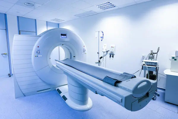
A CT scan is excellent for providing a broad, overall view of the abdomen and chest. It's great for spotting large tumours or distant spread of cancer. However, EUS provides far superior detail for small structures and allows for immediate biopsy, which a CT cannot do.
EUS vs. MRI: Complementary, Not Competitive
An MRI offers excellent soft-tissue contrast and is very good for imaging the liver and bile ducts (with MRCP). EUS and MRI are often used together. An MRI might identify a suspicious area in the pancreas, and EUS is then used to get a closer look and obtain a biopsy to confirm the diagnosis.
EUS vs. Standard Endoscopy: Seeing Beneath the Surface
A standard endoscopy is limited to the surface mucosa. It can see ulcers, inflammation, and polyps. EUS is the logical next step when a deeper view is needed—to see the layers beneath a lesion or to examine organs adjacent to the stomach and oesophagus.
Conclusion
Navigating a complex health concern requires not just expert care but also the most effective diagnostic tools. Endoscopic Ultrasound (EUS) represents a significant leap forward in medical imaging, offering a pathway to a fast, precise, and minimally invasive diagnosis. By providing unparalleled detail and the unique ability to combine diagnosis and biopsy in one session, EUS reduces the waiting and wondering that can be so taxing for any patient.
If your doctor suggests an EUS, it is likely because they are seeking the clearest possible picture to guide your treatment. Being informed about the procedure empowers you to ask the right questions and actively participate in your healthcare. Remember, a timely and accurate diagnosis is the most critical first step toward effective treatment and recovery.
Consult a Gastroenterologist for the best advice
Consult a Gastroenterologist for the best advice

Dr Rohit Sureka
Gastroenterology/gi Medicine Specialist
15 Years • MBBS, DNB General Medicine, DNB Gastroenterology
Jaipur
Apollo 247 virtual - Rajasthan, Jaipur

Dr. Umakanth Eskala
Gastroenterology/gi Medicine Specialist
16 Years • DM (GASTRO)
Visakhapatnam
Apollo 24|7 Clinic - Andhra Pradesh, Visakhapatnam

Dr Harish K C
Gastroenterology/gi Medicine Specialist
15 Years • MBBS MD DM MRCP(UK) (SCE-Gastroenterology and Hepatology)
Bangalore
Manipal Hospital, Bangalore

Dr. Paramesh K N
Gastroenterology/gi Medicine Specialist
16 Years • MBBS, MS ( General Surgery), DNB ( Surgical Gastroenterology)
Hyderabad
Sprint Diagnostics Centre, Hyderabad
Dr. Vijay Rai
Gastroenterology/gi Medicine Specialist
19 Years • MBBS,MD General Medicine,MD GASTROENTOLOGY
Kolkata
Livgastro, Kolkata
Consult a Gastroenterologist for the best advice

Dr Rohit Sureka
Gastroenterology/gi Medicine Specialist
15 Years • MBBS, DNB General Medicine, DNB Gastroenterology
Jaipur
Apollo 247 virtual - Rajasthan, Jaipur

Dr. Umakanth Eskala
Gastroenterology/gi Medicine Specialist
16 Years • DM (GASTRO)
Visakhapatnam
Apollo 24|7 Clinic - Andhra Pradesh, Visakhapatnam

Dr Harish K C
Gastroenterology/gi Medicine Specialist
15 Years • MBBS MD DM MRCP(UK) (SCE-Gastroenterology and Hepatology)
Bangalore
Manipal Hospital, Bangalore

Dr. Paramesh K N
Gastroenterology/gi Medicine Specialist
16 Years • MBBS, MS ( General Surgery), DNB ( Surgical Gastroenterology)
Hyderabad
Sprint Diagnostics Centre, Hyderabad
Dr. Vijay Rai
Gastroenterology/gi Medicine Specialist
19 Years • MBBS,MD General Medicine,MD GASTROENTOLOGY
Kolkata
Livgastro, Kolkata
Frequently Asked Questions
Is the EUS procedure painful?
No, the procedure is not painful. You will be under sedation (conscious sedation or sometimes deeper sedation) to ensure you are comfortable and relaxed. You will likely sleep through the entire procedure and have little to no memory of it.
How long does it take to get EUS biopsy results?
The procedure itself takes 30–60 minutes. However, the tissue samples obtained via biopsy need to be processed and analysed by a pathologist. This typically takes 3 to 7 working days. Your doctor will schedule a follow-up appointment to discuss the full results.
What are the risks of an EUS procedure?
EUS is considered very safe, with a low complication rate similar to that of a standard endoscopy. Potential risks include bleeding, infection, or perforation (a tear in the GI tract wall), but these are rare. The risk is slightly higher if a biopsy (FNA/FNB) is performed.
Is EUS better than a CT scan?
It's not about being universally 'better,' but about being the right tool for the job. A CT scan gives a broad overview of the entire abdomen. EUS provides exquisite detail of specific areas adjacent to the stomach and oesophagus. They are often used together to get a complete picture.
Can EUS be used for treatment?
Yes! This is known as interventional EUS. Beyond diagnosis, EUS can be used to drain pancreatic pseudocysts, place drains into the bile or pancreatic ducts, inject pain medication for chronic pancreatitis, and even perform tumour ablations in certain cases.


.webp)

