
PET-CT Scan: Purpose, Process, and Results
Learn about PET-CT scans, their purpose, how the procedure is done, and what the results mean for diagnosis and treatment planning.

Written by Dr. M L Ezhilarasan
Reviewed by Dr. D Bhanu Prakash MBBS, AFIH, Advanced certificate in critical care medicine, Fellowship in critical care medicine
Last updated on 24th Sep, 2025

Introduction
Facing a medical test can be daunting, especially when it has a complex name like a PET-CT scan. If your doctor has recommended one, you likely have many questions. This guide is designed to demystify the entire process, from the science behind the scan to what you can expect on the day and how to understand your results. We’ll walk you through the clinical practice of PET-CT imaging in clear, simple language, empowering you to be an active participant in your healthcare journey. Whether this scan is for investigating cancer, a heart condition, or a neurological issue, understanding the purpose, preparation, and procedure will help you feel more confident and prepared.
What is a PET-CT Scan, Really?
A PET-CT scan is a powerful imaging tool that combines two different technologies to give doctors a highly detailed picture of what’s happening inside your body. It’s more than just a simple X-ray; it provides both functional and structural information in a single session.
The PET Scan: Seeing Cellular Activity
The PET (Positron Emission Tomography) part of the scan focuses on function. Before the scan, a small amount of a radioactive sugar molecule, called a radiotracer (most commonly FDG), is injected into your bloodstream. Because cancer cells and some other types of diseased cells are often more active than normal cells, they absorb more of this radioactive sugar. The PET scanner detects the radiation emitted by these areas, creating colour-coded images that highlight regions of high metabolic activity. Think of it as a map showing which cells are "working overtime."
The CT Scan: Providing the Anatomical Map
The CT (Computed Tomography) part is like a very detailed, three-dimensional X-ray. It takes a series of images from different angles to create a cross-sectional view of your bones, organs, and tissues. This provides the anatomical "roadmap," showing the exact size, shape, and location of structures within the body.
Why Combining PET and CT is a Game-Changer
Before PET-CT, doctors had to perform separate scans and then mentally fuse the images, which could lead to uncertainty. The fusion of PET and CT in one machine is the true breakthrough. It precisely overlays the PET’s "activity map" onto the CT’s "anatomical map." This allows a radiologist to pinpoint exactly where abnormal activity is occurring. For example, they can see not just that there’s high metabolic activity in the chest, but that it’s specifically in a lymph node near the lung. This accuracy is crucial for effective diagnosis and treatment planning.
Consult a Radiologist for the best advice
Why Would Your Doctor Recommend a PET-CT Scan?
The most common reason for a PET-CT scan is in the field of oncology, but its applications extend to other areas of medicine as well.
In Oncology: The Primary Use Case
In cancer care, PET-CT imaging is an indispensable tool used throughout the patient journey.
• For Cancer Diagnosis and Staging: If a mass is found, a PET-CT can help determine if it’s cancerous based on its metabolic activity. More importantly, it is the gold standard for "staging" cancer, determining if and where the cancer has spread (metastasised) to lymph nodes or other organs. This staging directly influences the treatment approach.
• To Check Treatment Effectiveness: During or after treatment (like chemotherapy or radiation), a follow-up PET-CT scan can show if the therapy is working. A decrease in metabolic activity in the tumours is a positive sign that the cancer cells are responding.
• For Detecting Cancer Recurrence: After treatment is complete, PET-CT scans are used for surveillance to check for the return of cancer.
Beyond Cancer: Neurological and Cardiac Applications
While less common, PET-CT scans are also used to evaluate brain disorders like Alzheimer's disease (by detecting patterns of low metabolism) and to assess blood flow to the heart muscle in patients with coronary artery disease, helping to determine if heart muscle is still viable after a heart attack.
Preparing for Your PET-CT Scan: A Step-by-Step Guide
Proper preparation is critical for obtaining accurate results. The goal is to ensure your body’s cells, particularly muscle cells, aren't using too much sugar, which would make it harder to distinguish them from potentially diseased cells.
The Crucial Pre-Scan Diet and Fasting
You will be instructed to fast for typically 4-6 hours before your appointment. This means no food, candy, or sugary drinks. Water is usually encouraged to ensure you are well-hydrated. Strenuous exercise should be avoided for 24 hours before the scan, as it can alter muscle glucose uptake.
Managing Diabetes and Blood Sugar Levels
For diabetic patients, special instructions are essential. High blood sugar levels can interfere with the radiotracer’s absorption. You will receive specific guidance from your doctor on how to manage your medications (like insulin) and how to check your blood sugar before the scan. If you have diabetes, it is crucial to inform the imaging centre when you schedule your appointment.
What to Wear and What to Leave at Home
Wear comfortable, loose-fitting clothing without metal zippers or snaps. You will likely be asked to change into a gown. Leave jewellery and other metal accessories at home.
What to Expect on the Day of the Procedure
Knowing the steps can significantly reduce anxiety. The entire process usually takes between 2 to 3 hours.
Step 1: The Tracer Injection and Uptake Period
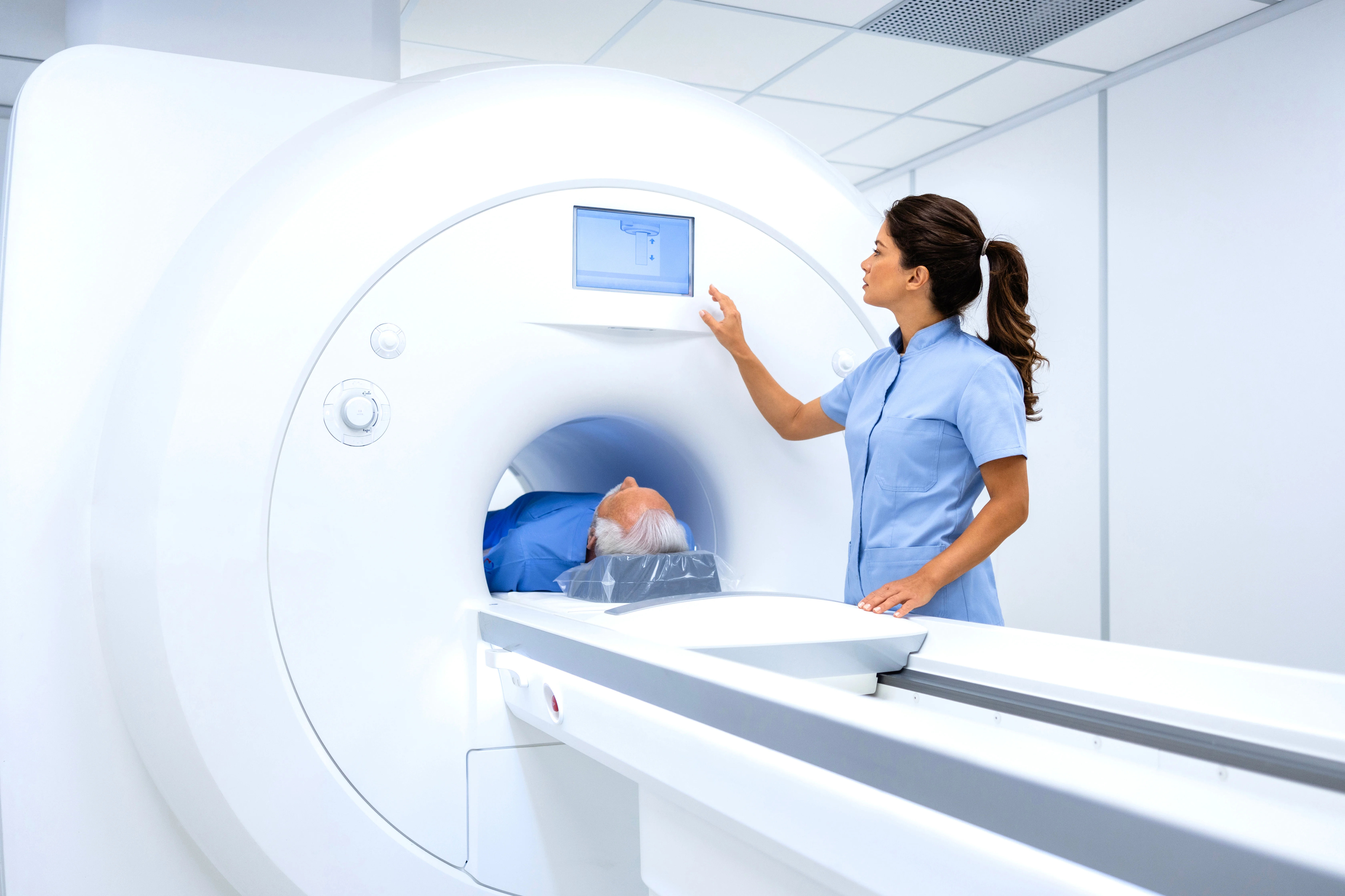
Upon arrival, a healthcare professional will insert an intravenous (IV) line, usually in your arm, to inject the radioactive tracer. This is a painless procedure similar to a blood draw. After the injection, you will rest quietly for about 60-90 minutes in a comfortable recliner. This "uptake period" allows the tracer to circulate and be absorbed by your body’s cells. You will be asked to relax and avoid moving or talking too much, as this can activate muscles and affect the images.
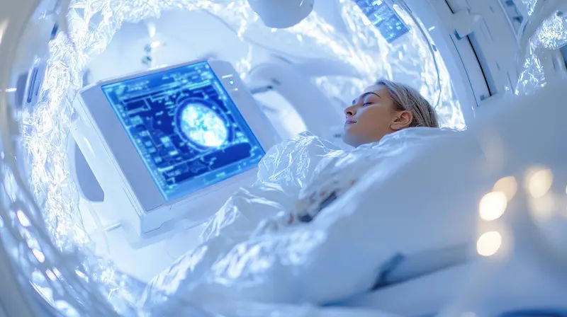
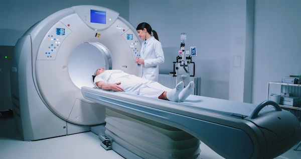
Step 2: The Scanning Process Itself
After the uptake period, you will be taken to the scanning room. The PET-CT machine looks like a large doughnut. You will lie on a padded table that slowly moves you through the scanner. It’s important to lie as still as possible during the scan, which typically lasts 20 to 45 minutes. The technologist will be in an adjacent room but can see and hear you at all times. You will be able to communicate with them through an intercom.
Understanding Your PET-CT Scan Results
You will not receive results immediately. The images must be analysed by a specialised radiologist who will prepare a detailed report for your referring doctor.
What Does a "Uptake" or "Hot Spot" Mean?
Areas of high radiotracer concentration appear as "hot spots" on the images. While this often indicates disease, it’s not always cancer. Inflammation, infection, or even normal muscle activity after exercise can also cause uptake. The radiologist uses their expertise to distinguish between these possibilities based on the location, pattern, and intensity (often measured as an SUV value) of the uptake.
The Radiologist's Report and Your Doctor's Interpretation
Your primary doctor or specialist is the best person to explain the results. They will combine the PET-CT findings with your medical history, physical exam, and other test results to give you a complete picture. They can tell you what the findings mean for your specific situation and next steps. It is essential to discuss the results directly with your doctor.
Safety, Risks, and Radiation Exposure
It's natural to be concerned about radiation. The radiation exposure from a PET-CT scan is considered safe for diagnostic purposes, but it is higher than a standard X-ray.
The Low-Dose Radiation Explained
The amount of radiation from the radiotracer is small and decays quickly. The CT component also contributes to the total dose. However, the medical benefit of obtaining an accurate diagnosis far outweighs the potential risk from radiation exposure. The principles of radiation safety (using the minimum necessary dose) are always followed.
Special Considerations: Pregnancy and Breastfeeding
If you are pregnant or suspect you might be, you must inform your doctor and the imaging centre immediately. The scan will generally be avoided unless absolutely critical. If you are breastfeeding, you will be given specific instructions, which may include pumping and discarding breast milk for a short period after the scan.
Conclusion: An Empowered Approach to Your Health
A PET-CT scan is a sophisticated and invaluable tool in modern medicine. While the idea of undergoing such a procedure can be intimidating, understanding its purpose, the meticulous preparation involved, and what happens during the scan can alleviate much of the anxiety. Remember, this scan provides your medical team with critical information to make the best possible decisions about your care. By asking informed questions and following pre-scan instructions carefully, you become an active partner in your health journey. The goal is always to obtain clear, accurate information to guide effective treatment and, ultimately, improve health outcomes. If you have any lingering concerns after discussing the scan with your doctor, don't hesitate to seek a second opinion or ask for further clarification.
Consult a Radiologist for the best advice
Consult a Radiologist for the best advice

Dr.priyank Ks Chaudhary
Radiologist
10 Years • MBBS DMRD & DNB
Lucknow
Apollo Clinic Hazratganj, Lucknow

Dr. Surendra K
Radiologist
29 Years • MBBS, MD (Radio-Diagnosis)
Bengaluru
Apollo Clinic, Koramangala, Bengaluru
Dr. Karthik H V
Radiologist
2 Years • MBBS MD(Radio-Diagnosis),DNB
Mysuru
Apollo Clinic, Mysore, Mysuru

Dr. Gandham Sohini
Radiologist
3 Years • MBBS,MD(Radiology)
Manikonda Jagir
Apollo Clinic, Manikonda, Manikonda Jagir
Dr.kiran Barla
Radiologist
5 Years • MBBS, DNB (Radiology)
Hanamkonda
Apollo Clinic Hanamkonda, Hanamkonda
Consult a Radiologist for the best advice
Dr.priyank Ks Chaudhary
Radiologist
10 Years • MBBS DMRD & DNB
Lucknow
Apollo Clinic Hazratganj, Lucknow
Dr. Surendra K
Radiologist
29 Years • MBBS, MD (Radio-Diagnosis)
Bengaluru
Apollo Clinic, Koramangala, Bengaluru
Dr. Karthik H V
Radiologist
2 Years • MBBS MD(Radio-Diagnosis),DNB
Mysuru
Apollo Clinic, Mysore, Mysuru
Dr. Gandham Sohini
Radiologist
3 Years • MBBS,MD(Radiology)
Manikonda Jagir
Apollo Clinic, Manikonda, Manikonda Jagir
Dr.kiran Barla
Radiologist
5 Years • MBBS, DNB (Radiology)
Hanamkonda
Apollo Clinic Hanamkonda, Hanamkonda
.webp)
Frequently Asked Questions
1. How is a PET-CT scan different from an MRI?
A PET-CT scan shows metabolic activity (function), while an MRI provides extremely detailed images of soft tissues (structure) without using radiation. They are often used for different purposes; your doctor will choose the best tool for your specific condition.
2. What does it mean if my PET-CT scan is 'clear'?
A 'clear' or negative scan typically means no abnormal areas of high metabolic activity were detected that would suggest active disease, such as cancer. This is generally very good news, but your doctor will interpret this result in the context of your overall health.
3. Can I be around my family after a PET-CT scan?
The radiation from the tracer decreases rapidly. The general advice is to avoid prolonged close contact with infants and pregnant women for a few hours after the scan. The imaging staff will give you specific instructions before you leave.
4. Is the radiotracer used in a PET-CT scan safe?
Yes, the radiotracer is FDA-approved and considered safe. Allergic reactions are extremely rare. It leaves your body primarily through urine within a few hours to a day.
5. What is an SUV value on my PET-CT report?
SUV (Standardised Uptake Value) is a semi-quantitative measurement of how much radiotracer is concentrated in a tissue. A higher SUV generally indicates more metabolic activity. However, only a trained radiologist can determine if a high SUV is significant based on its location and other factors.