
ECG vs. Echo; Understanding Your Heart Tests
Confused about ECG vs. Echo? Learn the key differences between these common heart tests, what each detects, how they’re done, and why your doctor might order one or both to assess your heart health.

Written by Dr. Siri Nallapu
Reviewed by Dr. Shaik Abdul Kalam MD (Physician)
Last updated on 5th Sep, 2025

When your doctor wants to check your heart health, two of the most common tests they might order are an electrocardiogram (ECG or EKG) and an echocardiogram (Echo). While their names sound similar and both are crucial for cardiac care, they serve very different purposes. Many patients are unsure about the difference between ECG and Echo, often confusing what each test reveals. This guide will demystify these essential diagnostic tools, explaining how they work, what they look for, and why your doctor might choose one over the other—or even both. Understanding these tests can empower you to take an active role in your heart health journey.
What is an Electrocardiogram (ECG or EKG)?
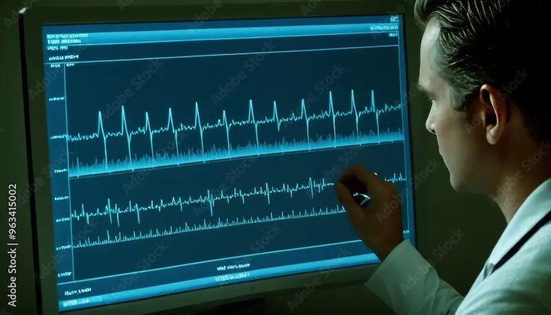
An electrocardiogram, commonly known as an ECG or EKG, is a quick, non-invasive test that measures the electrical activity of your heart. Think of it as a "speedometer" for your heart's rhythm. It provides a snapshot of the heart's electrical impulses as they travel through the heart muscle, causing it to contract (beat).
How Does an ECG Work?
During an ECG, small, sticky electrodes are attached to your chest, arms, and legs. These sensors detect the tiny electrical signals produced each time your heart beats. A machine records these signals and prints them out as waves on a graph. The entire process is painless and takes only a few minutes. There's even a portable version called a Holter monitor that records your heart's activity over 24 or 48 hours to catch irregular rhythms that might not appear during a brief in-office test.
What Does an ECG Detect?
The primary function of an ECG is to assess the heart's rhythm and rate. It is the gold standard for identifying:
- Arrhythmias: Irregular heartbeats, such as atrial fibrillation or tachycardia.
- Heart Attacks: It can show evidence of a current or previous heart attack by revealing areas of damaged heart tissue.
- Coronary Artery Disease: Signs of reduced blood flow to the heart muscle.
- Structural Problems: Certain issues, like an enlarged heart or thickened heart muscle, can sometimes be inferred.
What is an Echocardiogram (Echo)?
An echocardiogram, or Echo, is an ultrasound of the heart. If an ECG is the heart's speedometer, an Echo is its video camera. It uses high-frequency sound waves (ultrasound) to create detailed, moving images of your heart's structures, valves, and blood flow in real-time.
How Does an Echo Work?
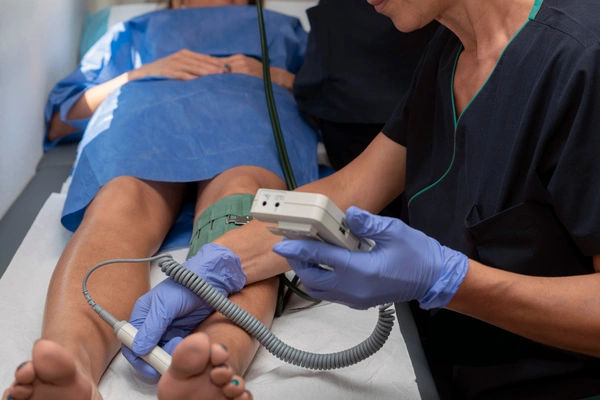
A technician, called a sonographer, applies a special gel to your chest and then uses a handheld device called a transducer. The transducer emits sound waves that bounce off your heart structures. These returning "echoes" are translated by a computer into live images on a screen. This is known as a transthoracic echocardiogram, the most common type. Other types include a stress echocardiogram (images taken after exercise) and a transesophageal echo (TEE), where a probe is guided down the esophagus for even clearer images.
Consult a Specialist for Personalised Advice
What Does an Echo Detect?
An Echo provides a comprehensive look at the heart's anatomy and function. It is excellent for diagnosing:
- Heart Valve Disease: Assessing leaky or narrowed valves.
- Pumping Function: Precisely measuring the heart's ejection fraction, which is how well it pumps blood.
- Heart Muscle Problems: Identifying cardiomyopathies (thickened or weakened heart muscle).
- Pericardial Disease: Fluid around the heart or inflammation of the sac surrounding the heart.
- Congenital Heart Defects: Structural problems present from birth.
- Blood Clots or Tumors: Detecting masses within the heart chambers.
Key Differences Between ECG and Echo; A Side-by-Side Comparison
Why Would a Doctor Order One Test Over the Other?
The choice between an ECG and an Echo depends entirely on the clinical question your doctor needs to answer.
- An ECG is typically ordered first when a patient presents with symptoms like chest pain, palpitations, dizziness, or shortness of breath. It's a fantastic screening tool to quickly rule out acute, life-threatening issues like a heart attack or a dangerous arrhythmia. It's also part of a routine physical, especially before surgery.
- An Echo is ordered when the initial ECG is abnormal, or when the symptoms suggest a problem with the heart's structure or pumping ability. For example, if a doctor hears a heart murmur (suggesting a valve problem) or if a patient has signs of heart failure like leg swelling and fatigue, an Echo is the logical next step to visualize the issue.
Often, the tests are complementary. An ECG might detect an irregular rhythm, and then an Echo is used to check if that rhythm problem has weakened the heart muscle.
The Patient Experience: What to Expect
During an ECG
You will lie down on an exam table. A nurse or technician will clean areas on your chest, arms, and legs and attach the electrodes. You'll need to lie still and breathe normally for about a minute while the machine records the signals. There is no sensation or pain; you only feel the sticky electrodes on your skin.
During an Echo
You will also lie on an exam table, usually on your left side. The sonographer will apply gel to your chest and press the transducer firmly against your skin, moving it to different angles. You may hear a "whooshing" sound—this is the machine amplifying the sound of blood flowing through your heart. The technician may ask you to hold your breath briefly to get clearer images. The gel might feel cool, but the test is entirely painless.
Interpreting the Results: What Do They Mean?
- ECG Results: A doctor looks at the pattern of the waves (P wave, QRS complex, T wave) for intervals, shapes, and rhythms that fall outside the normal range. An abnormal ECG might indicate anything from a harmless minor variation to a serious block in electrical conduction.
- Echo Results: A cardiologist analyzes the moving images to measure chamber sizes, wall thickness, valve motion, and pumping strength. A report will detail findings like "ejection fraction of 55-60% (normal)" or "mild mitral valve regurgitation."
It is crucial to discuss your results with your doctor, who can interpret them in the context of your overall health and symptoms.
Conclusion
In the world of cardiology, the ECG and echocardiogram are fundamental tools that, while different, work together to provide a holistic view of your heart's health. One isn't "better" than the other; they simply answer different questions. The ECG excels as a rapid-assessment tool for the heart's electrical system, while the Echo provides a deep dive into its physical structure and mechanical function. If your doctor recommends either, or both, of these tests, it's a proactive step towards understanding and safeguarding your most vital organ. Always feel empowered to ask your healthcare provider why a specific test is being ordered and what they hope to learn from it. Your heart health is a partnership, and understanding these diagnostics is a key part of that journey.
Consult a Specialist for Personalised Advice
Consult a Specialist for Personalised Advice

Dr. Vasanthasree Nair
General Practitioner
15 Years • MBBS
Angamaly
Apollo 24|7 Clinic - Kerala, Angamaly
(575+ Patients)

Dr. Pallavi Nedurumalli Reddy
General Practitioner
9 Years • MBBS
Hyderabad
Apollo 24|7 Clinic - Telangana, Hyderabad
(25+ Patients)

Dr. Praveen Kumar Mukka
General Physician/ Internal Medicine Specialist
21 Years • MBBS, MD General Medicine
Hyderabad
Apollo 24|7 Clinic - Telangana, Hyderabad
(150+ Patients)
Dr. Musaddiqa Khanum
General Practitioner
18 Years • MBBS, DNHE MPH.
Hubli
Vishwas Clinic, Hubli
Dr.f.fathima Thahira Juhi
General Practitioner
6 Years • MBBS
Chennai
YZEE CLINIC AND DIAGNOSTICS, Chennai
Consult a Specialist for Personalised Advice
Dr. Vasanthasree Nair
General Practitioner
15 Years • MBBS
Angamaly
Apollo 24|7 Clinic - Kerala, Angamaly
(575+ Patients)
Dr. Pallavi Nedurumalli Reddy
General Practitioner
9 Years • MBBS
Hyderabad
Apollo 24|7 Clinic - Telangana, Hyderabad
(25+ Patients)
Dr. Praveen Kumar Mukka
General Physician/ Internal Medicine Specialist
21 Years • MBBS, MD General Medicine
Hyderabad
Apollo 24|7 Clinic - Telangana, Hyderabad
(150+ Patients)
Dr. Musaddiqa Khanum
General Practitioner
18 Years • MBBS, DNHE MPH.
Hubli
Vishwas Clinic, Hubli
Dr.f.fathima Thahira Juhi
General Practitioner
6 Years • MBBS
Chennai
YZEE CLINIC AND DIAGNOSTICS, Chennai
.webp)
.webp)
Frequently Asked Questions
Can an ECG detect a blocked artery?
An ECG can suggest reduced blood flow to the heart muscle, which may be caused by a blocked artery, especially during a heart attack. However, it is not a definitive test for blockages. A cardiac catheterization or CT coronary angiogram is typically used to directly visualize blocked arteries.
Which is more accurate, an ECG or an Echo?
This is like asking if a thermometer is more accurate than a blood pressure cuff—they measure different things. For rhythm problems, an ECG is more direct and accurate. For assessing structural issues like valve function or pumping strength, the Echo is unequivocally more accurate and detailed.
Why would I need an Echo if my ECG is normal?
A normal ECG only means your heart's electrical activity and rhythm are normal at that moment. It does not rule out structural problems. If you have symptoms like shortness of breath or a heart murmur, an Echo is necessary to check for issues like weak heart muscle or leaky valves that an ECG cannot see.
Do I need to prepare for an ECG or Echo?
For a standard ECG, no special preparation is needed. For a standard transthoracic Echo, you also typically do not need to prepare. However, for a transesophageal echo (TEE), you will need to fast (not eat or drink) for several hours beforehand as it involves sedation.
How often should you get a routine ECG or Echo?
There is no standard guideline for the general public. These tests are ordered based on symptoms, risk factors (like high blood pressure, diabetes, or family history), or as follow-up for a known heart condition. You should discuss the need for routine screening with your doctor.