Glaucoma: Causes, Symptoms and Diagnosis
Understand glaucoma with insights into its causes, symptoms, and diagnosis. Learn how early detection can help protect your vision and eye health.

Written by Dr. Siri Nallapu
Reviewed by Dr. Dhankecha Mayank Dineshbhai MBBS
Last updated on 13th Jan, 2026

Introduction
Glaucoma is a leading cause of irreversible blindness worldwide, yet it often progresses silently, without any early warning signs. This group of eye diseases damages the optic nerve, typically due to elevated pressure inside the eye, and gradually steals your peripheral vision. Because this vision loss is permanent, understanding what leads to glaucoma and how it is diagnosed is your most powerful tool for prevention. This article will guide you through the causes and risk factors, help you recognise the subtle and urgent signs, and provide a detailed look at the diagnostic tests that can save your sight. Early glaucoma diagnosis through a comprehensive eye exam is the only way to detect the disease before significant damage occurs, allowing for treatment that can halt its progression.
What is Glaucoma? The Silent Thief of Sight
Glaucoma isn't a single disease but a group of conditions that result in optic neuropathy, damage to the optic nerve, which is the vital cable that transmits visual information from your eye to your brain. This damage creates blind spots in your visual field, often starting with your side (peripheral) vision.
How Glaucoma Damages Your Vision
The most common cause of this damage is high intraocular pressure (IOP). Inside your eye, a clear fluid called aqueous humor circulates to nourish eye tissues. It drains out through a tiny meshwork. If this drainage system becomes inefficient, fluid builds up, pressure rises, and this increased IOP can compress and damage the delicate fibers of the optic nerve.
Primary Open-Angle Glaucoma vs. Acute Angle-Closure Glaucoma
Primary Open-Angle Glaucoma (POAG): This is the most common form, accounting for at least 90% of cases. The "angle" where fluid drains is open but becomes clogged over time, like a slow-draining sink. Pressure builds gradually, causing no pain and initially no vision changes.
Acute Angle-Closure Glaucoma: This is a medical emergency. The angle between the iris and cornea is too narrow, and if it becomes completely blocked, eye pressure can rise very suddenly. This causes severe pain, nausea, and blurred vision and requires immediate treatment to prevent rapid vision loss.
Consult an Ophthalmologist for the best advice
What Leads to Glaucoma? Understanding the Causes and Risk Factors
While high eye pressure is a major factor, it's not the only one. Some people with normal IOP can develop glaucoma (normal-tension glaucoma), while others with high IOP may never do so. It's a combination of factors that determines your risk.
The Primary Culprit: High Intraocular Pressure (IOP)
Elevated IOP is the most significant and only modifiable risk factor. It's not the cause itself but the primary mechanism that leads to optic nerve damage in most individuals.
Key Risk Factors You Can't Control
Age and Family History: Your risk increases significantly after age 60. If you have a parent or sibling with glaucoma, your risk is much higher, indicating a possible genetic link.
Ethnicity and Corneal Thickness: People of African, Asian, and Hispanic descent are at increased risk for certain types of glaucoma. Additionally, having a thinner cornea can make you more susceptible to optic nerve damage from IOP.
Medical Conditions That Increase Your Risk
Diabetes, Hypertension, and Heart Disease: These systemic conditions can affect blood flow to the optic nerve, increasing glaucoma risk. If you have been diagnosed with any of these, it's crucial to inform your eye doctor. Managing chronic conditions like diabetes is key; if your symptoms are difficult to control, consulting a specialist online with Apollo24|7 can help you create an effective management plan.
The Signs and Symptoms: What to Watch For
The symptoms of glaucoma depend entirely on the type and stage of the disease.
The Deceptive Silence of Open-Angle Glaucoma
This is why POAG is so dangerous. In its early stages, there are no symptoms. No pain, no redness, and your vision seems perfectly clear. The damage begins in your peripheral vision, which your brain compensates for until the loss is severe.
The Urgent Signs of Acute Angle-Closure Glaucoma
An attack is a medical emergency. Symptoms appear suddenly and include:
Severe eye pain and headache
Nausea and vomiting
Seeing halos or rainbows around lights
Sudden blurred vision
Redness in the eye
If you experience these symptoms of angle-closure glaucoma, seek immediate medical care.
Later-Stage Symptoms: When Vision Loss Becomes Apparent
As all forms of glaucoma progress, you may notice:
Gradual loss of peripheral vision, often described as "tunnel vision."
Blurred or patchy blind spots in your side vision.
In advanced stages, difficulty seeing in low light and a loss of central vision.
How Glaucoma is Diagnosed: A Step-by-Step Guide to the Tests
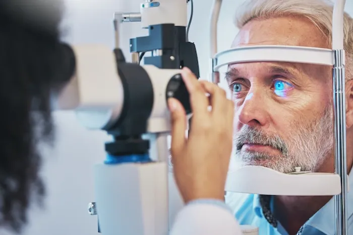
A glaucoma diagnosis is not based on a single test. It requires a comprehensive dilated eye exam where an ophthalmologist pieces together evidence from several procedures.
The Comprehensive Dilated Eye Exam: Your First Defense
This is the cornerstone of detection. If you're over 40 or have risk factors, booking a comprehensive eye exam is the most important step you can take.
Tonometry: Measuring Eye Pressure
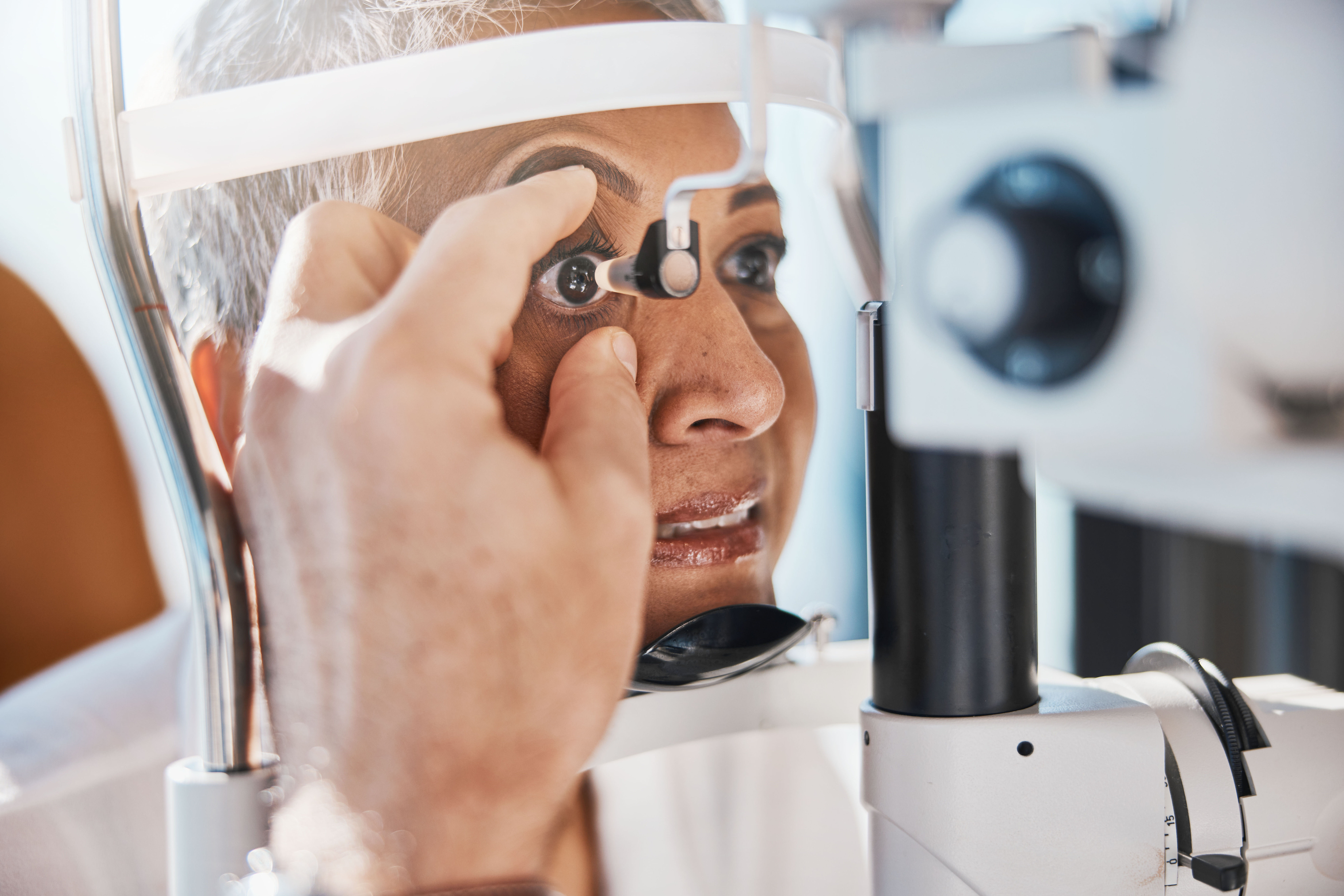
This test measures your intraocular pressure (IOP). The most common method is the "air puff" test (non-contact tonometry), but a more accurate method involves a device that gently touches the eye's surface after it's numbed with drops (applanation tonometry).
Ophthalmoscopy and Slit-Lamp Exam: Viewing the Optic Nerve
After dilating your pupils with eye drops, your doctor uses a special magnifying lens (ophthalmoscope) and a slit-lamp microscope to examine the shape and color of your optic nerve. They look for signs of damage, such as cupping, where the nerve's central area appears to become larger and hollowed out.
Perimetry (Visual Field Test): Checking Your Side Vision
This computerised test maps your complete field of vision. You'll be asked to press a button whenever you see a light spot appear in your peripheral vision. It creates a detailed map to identify any blind spots (scotomas) caused by glaucoma.
Gonioscopy: Examining the Drainage Angle
A special contact lens with a mirror is placed on the eye to examine the drainage angle where the iris meets the cornea. This critical test determines whether the angle is open (as in POAG) or narrow/closed (as in angle-closure glaucoma).
Pachymetry: Measuring Corneal Thickness
This simple, painless test uses a small probe to measure the thickness of your cornea. A thinner cornea can sometimes give an artificially low IOP reading, while a thicker cornea can give an artificially high one. This measurement helps your doctor interpret your tonometry results more accurately.
Optical Coherence Tomography (OCT): A High-Tech Scan
This advanced imaging technology provides high-resolution, cross-sectional images of the retina and optic nerve. It can measure the thickness of the nerve fiber layer, allowing doctors to detect damage often years before it becomes visible on a traditional exam or causes vision loss on a visual field test.
Who Should Get Screened and How Often?
The American Academy of Ophthalmology recommends a comprehensive eye exam:
Before age 40: Every 5-10 years.
Ages 40-54: Every 2-4 years.
Ages 55-64: Every 1-3 years.
Age 65 and older: Every 1-2 years.
You need more frequent screening if you are in a high-risk group (e.g., family history, African or Hispanic heritage, previous eye injury, diabetes). In these cases, your doctor will recommend a schedule, which may be annually.
Conclusion
Understanding the pathways that lead to glaucoma from high eye pressure and genetic predisposition to underlying health conditions empowers you to take proactive steps for your eye health. Recognising that the most common form is symptomless until significant damage has occurred underscores the non-negotiable importance of regular, comprehensive eye exams. The diagnostic process is thorough and painless, designed to catch the slightest hint of the disease long before it affects your quality of life. If you are overdue for an eye exam or fall into a high-risk category, do not wait for symptoms to appear. Protecting your vision is a lifelong commitment, and it starts with a single appointment.
Consult an Ophthalmologist for the best advice
Consult an Ophthalmologist for the best advice

Dr Ranojit Basu
Ophthalmologist
24 Years • MBBS, DNB Ophthalmology, Diploma in Ophthalmic Medicine and. Surgery
Kolkata
Titanium Eye Care, Kolkata

Dr. Atheeshwar Das
Ophthalmologist
15 Years • MBBS,DO,DNB(Gold Medal),FRCS(Glasgow),FICO(UK),
Chennai
Apollo Speciality Hospitals OMR, Chennai

Dr. Sunanda Nandi
Ophthalmologist
7 Years • MBBS, MS (Ophthalmology)
Silchar
Apollo Clinic Silchar, Silchar

Dr. Sridhar Annam
Ophthalmologist
15 Years • MBBS, DO, MS (Opthal)
Hyderabad
Apollo Hospitals Jubilee Hills, Hyderabad
(25+ Patients)
Meghana Kotesh
Ophthalmologist
3 Years • MBBS, MS (OPTHALMOLOGIST )
Bengaluru
Apollo Medical Center, Marathahalli, Bengaluru
More articles from Glaucoma
Frequently Asked Questions
1. Can you test for glaucoma at home?
No, you cannot reliably test for glaucoma at home. While monitoring your overall vision is good practice, the key indicators of glaucoma, like elevated eye pressure, optic nerve damage, and peripheral vision loss, require specialized medical equipment and a trained ophthalmologist for accurate diagnosis.
2. What are the first signs of glaucoma that you might notice?
For the most common type (open-angle glaucoma), there are typically no early signs you would notice. The first noticeable change is often a loss of peripheral or side vision, but this only occurs after significant, irreversible damage has already happened. This is why screening exams are so critical.
3. Is glaucoma curable once diagnosed?
There is currently no cure for glaucoma, and any vision loss that has occurred cannot be reversed. However, it is highly treatable. With early glaucoma diagnosis and consistent treatment (usually medicated eye drops, laser therapy, or surgery), the progression of the disease can almost always be halted, preserving your remaining vision.
4. What is the main cause of glaucoma?
The primary mechanism is damage to the optic nerve, most commonly caused by elevated pressure inside the eye (intraocular pressure). This pressure builds up when the eye's natural drainage system for aqueous humor fluid becomes inefficient.
5. How long does it take to go blind from glaucoma without treatment?
The timeline varies greatly from person to person. Without treatment, open-angle glaucoma can take many years to lead to blindness. However, an acute angle-closure glaucoma attack can cause blindness within a day if not treated immediately. The unpredictable nature of the disease makes early intervention essential.


