
Glaucoma Diagnosis: Signs, Risks, and How Doctors Confirm It
Know about glaucoma, what it is, how it develops, risks, symptoms, causes, screening, prevention and living with glaucoma and more.

Written by Dr. Siri Nallapu
Reviewed by Dr. Rohinipriyanka Pondugula MBBS
Last updated on 13th Jan, 2026

Introduction
Glaucoma is a group of eye diseases that damage the optic nerve—the “data cable” that carries images from your eye to your brain. It’s one of the leading causes of irreversible vision loss worldwide, yet early glaucoma rarely has obvious symptoms. That’s why understanding the subtle signs, knowing your personal risk, and learning how the glaucoma diagnosis works can make the difference between preserving sight and losing it. In this guide, we break down exactly what leads to the signs of glaucoma and how doctors confirm a diagnosis. You’ll learn which symptoms need urgent attention, which tests are typically performed (like eye pressure checks, optic nerve imaging, and visual field testing), and why some people develop glaucoma even with “normal” eye pressure.
What Is Glaucoma? A Quick, Clear Definition
Glaucoma refers to a family of eye conditions that progressively damage the optic nerve, usually in the context of problems with fluid drainage in the eye. The optic nerve is made up of more than a million retinal ganglion cell axons. Once these cells are damaged, vision loss is permanent. Early on, glaucoma affects peripheral vision; without treatment, it can constrict your visual field and eventually impact central vision.
Consult a Top Ophthalmologist for Personalised Advice
The main types include:
- Primary open-angle glaucoma (POAG): The most common type, especially in adults. The eye’s drainage system (trabecular meshwork) becomes less efficient over time, often leading to elevated intraocular pressure (IOP) and gradual optic nerve damage. Early symptoms are rare—most people don’t notice changes until damage is advanced.
- Angle-closure glaucoma (also called closed-angle): The drainage angle becomes blocked, which can rapidly raise eye pressure. This may cause sudden eye pain, blurred vision, halos around lights, headache, and nausea. This is an emergency requiring same-day treatment.
- Normal-tension glaucoma: Optic nerve damage occurs even when measured eye pressure is in the “normal” range. Factors such as optic nerve susceptibility and vascular issues may contribute.
- Secondary glaucomas: Caused by another condition (e.g., steroid use, inflammation, trauma, pigment dispersion, pseudoexfoliation) that impairs fluid drainage or otherwise harms the optic nerve.
How Glaucoma Develops: Pressure, Outflow, and the Optic Nerve
Inside your eye, clear fluid called aqueous humour nourishes tissues and maintains shape. It’s continuously produced and drains through the trabecular meshwork into Schlemm’s canal. In open-angle glaucoma, this outflow pathway becomes less efficient, and pressure may rise. Over time, elevated IOP can compress and damage the optic nerve head, particularly where retinal ganglion cell axons converge (the lamina cribrosa).
But glaucoma biology goes beyond pressure:
- Mechanical stress: Sustained or fluctuating IOP can strain the optic nerve head and supporting tissues. This contributes to the characteristic “cupping” seen during a dilated exam.
- Vascular factors: Reduced blood flow, blood pressure dips at night, and dysregulation may make the optic nerve more vulnerable—even at “normal” pressures. This helps explain normal-tension glaucoma.
- Susceptibility differences: Thin corneas, genetic factors, and optic nerve anatomy vary from person to person, affecting risk and damage patterns.
Why “normal” pressure can still be harmful
Normal-tension glaucoma reminds us that “normal” is a population range, not a guarantee of safety. If your optic nerve is structurally vulnerable or blood flow is compromised (for example, with sleep apnea or extreme nocturnal blood pressure dips), even average pressures can be damaging. Conversely, some people with ocular hypertension (high pressure without nerve damage) may never develop glaucoma, but they need monitoring because the risk increases with higher IOP and thinner corneas.
Who Is at Risk? Major Risk Factors You Should Know
Some people are more likely to develop glaucoma and to progress faster if untreated:
- Age: Risk rises over 40 and increases with each decade.
- Family history: Having a first-degree relative with glaucoma greatly increases risk; this is particularly important for POAG.
- Ancestry: People of African and Afro-Caribbean descent have a higher risk and often an earlier onset; Hispanic/Latino individuals also face a higher risk as they age; certain Asian populations have an increased risk of angle-closure and normal-tension glaucoma.
- Elevated IOP and thin central cornea: Higher pressure is a strong risk factor; a thinner cornea not only raises risk but also can make pressure readings seem lower than they really are. The Ocular Hypertension Treatment Study identified thin corneas as a key risk factor for conversion to glaucoma.
- High myopia (nearsightedness): Linked to structural changes that can increase vulnerability.
Steroid use: Long-term steroid drops, pills, or injections can raise IOP in susceptible individuals.
Systemic health: Diabetes, sleep apnea, and vascular factors (very low nighttime blood pressure) may contribute to risk or faster progression.
If glaucoma or blindness runs in your family, schedule regular eye exams with dilation. People with diabetes should also keep metabolic health on track; if you need diabetes screening, Apollo 24|7 offers convenient home collection for tests like HbA1c and lipid panels. Keeping overall health in range supports eye health, too.
Early Signs and Symptoms: What You Might Notice (and What You Won’t)
Open-angle glaucoma often has no early warning signs. Vision loss starts in the periphery and is subtle. That’s why many people are unaware they have glaucoma until significant damage has occurred. Common early experiences include mild difficulty with side vision or occasional patchy areas—often noticed only on formal visual field testing.
Angle-closure glaucoma is different. It can present suddenly with:
- Severe eye pain and headache
- Blurred vision and halos around lights
- Red eye, nausea, and vomiting
- Mid-dilated, fixed pupil on exam
This is an emergency. Sudden symptoms like these require immediate care the same day to prevent permanent vision loss. If you experience these symptoms, seek emergency eye care. If you’re unsure, consult a doctor online with Apollo 24|7 for urgent guidance while arranging an in-person evaluation.
Normal-tension glaucoma may have non-specific visual complaints (like “washed out” contrast or patchy areas of vision) despite typical IOP readings. People may also notice glare or difficulty adjusting from light to dark. None of these symptoms are diagnostic on its own; diagnosis requires careful testing.
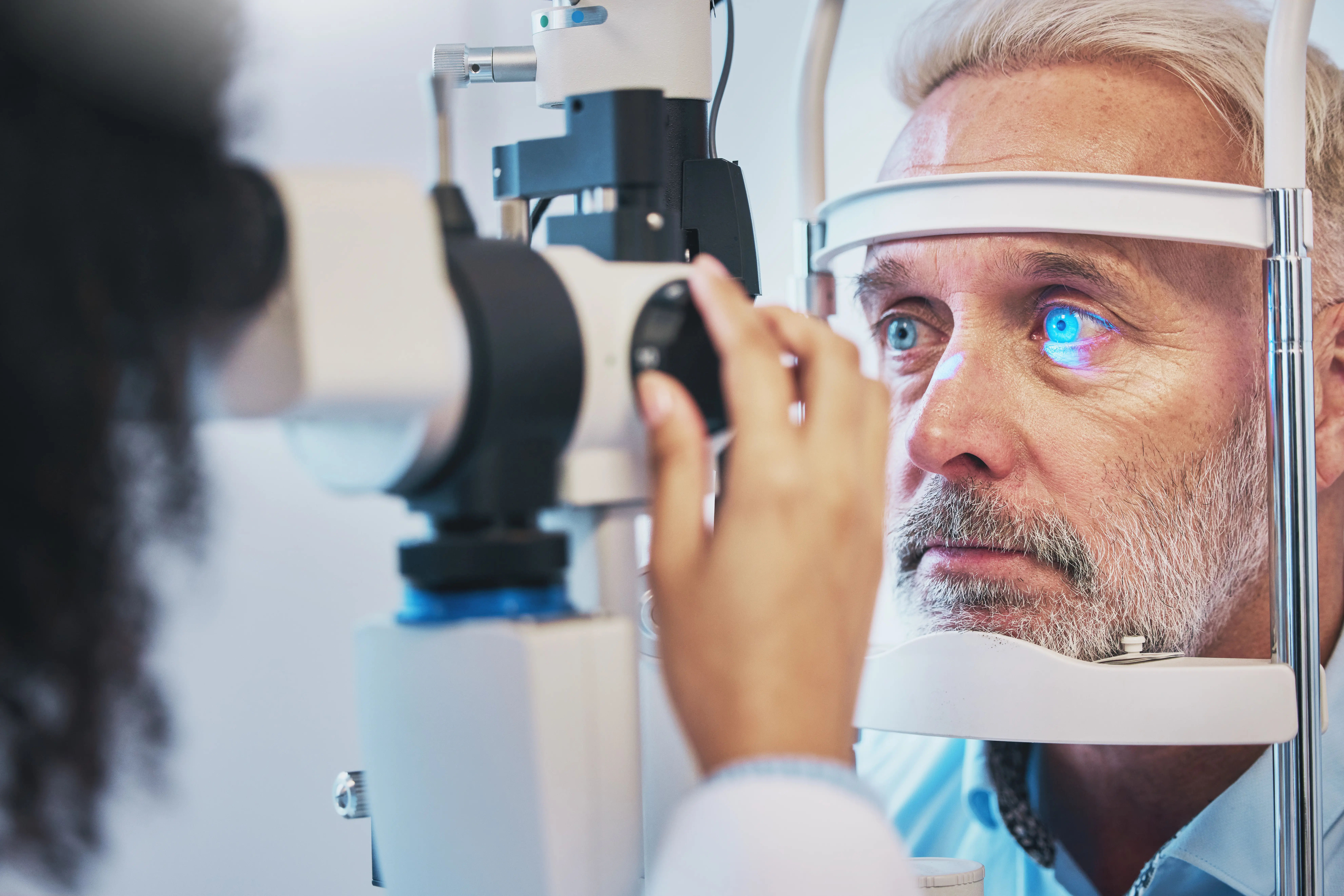
How Doctors Diagnose Glaucoma: Tests You Can Expect
There is no single test for glaucoma. Diagnosis is a synthesis of findings over time—pressures, optic nerve appearance, corneal thickness, drainage angle status, imaging, and functional tests. Expect several of the following:
- Tonometry (eye pressure measurement): Measures IOP, often with a blue light and a numbing drop (Goldmann applanation) or a handheld device. Normal ranges vary by person, and a single reading is not definitive. Pressures can fluctuate throughout the day. Pachymetry (central corneal thickness): A thinner cornea can make IOP readings appear artificially low and is itself a risk factor for developing glaucoma. This measurement helps your clinician interpret IOP and overall risk.
- Gonioscopy (examining the drainage angle): Using a special lens, your doctor checks whether your angle is open or narrow/closed, and looks for signs of secondary causes (e.g., pigment, pseudoexfoliation, neovascularisation).
- Dilated optic nerve evaluation: The clinician examines the optic nerve head for cupping, rim thinning, notching, disc haemorrhages, and asymmetry. Stereo photos may be taken for baseline comparison.
- OCT (optical coherence tomography): A painless imaging test that measures the retinal nerve fibre layer and ganglion cell complex thickness. OCT detects structural damage early and tracks change over time.
- Visual field testing (perimetry): Assesses functional vision—are there blind spots or areas of reduced sensitivity? Repeating fields confirms reliability and progression.
Special Cases, Look-Alikes, and When to Worry
Ocular hypertension vs glaucoma
- Ocular hypertension: Elevated IOP without optic nerve damage or visual field loss. Not everyone with ocular hypertension develops glaucoma, but the risk increases with higher IOP, thin corneas, and age. The Ocular Hypertension Treatment Study showed that lowering IOP reduces the risk of conversion to glaucoma.
- Glaucoma: Optic nerve damage and/or corresponding visual field defects. Treatment aims to reduce IOP and stabilise progression.
Secondary glaucomas
- Steroid-induced: From drops, injections, or systemic steroids; pressure can spike significantly.
- Pigmentary glaucoma: Pigment granules clog the trabecular meshwork, often in younger myopic adults.
- Pseudoexfoliation glaucoma: Flaky material deposits on lens and trabecular meshwork; common in older adults, may progress faster.
- Neovascular glaucoma: New, fragile blood vessels obstruct the angle; linked to diabetes or retinal vein occlusion; requires urgent speciality care.
Look-alikes and mimickers
- Optic neuropathies (ischemic, compressive) can mimic glaucoma field defects.
- High myopia can make the optic nerves look “glaucomatous.”
- Artefacts on OCT or unreliable visual fields can confuse the diagnosis.
If the diagnosis is unclear, your doctor may order repeat tests, further imaging, or refer you to a glaucoma specialist. If symptoms persist or vision problems worsen, consult a doctor online with Apollo24|7 for fast guidance and arrange an in-person exam promptly.
Screening, Prevention, and When to See a Doctor
Who should be screened?
- Adults over 40, especially with risk factors (family history, African or Hispanic/Latino ancestry, high myopia), should have regular comprehensive eye exams, including dilation.
- People with diabetes, long-term steroid use, or a history of eye injury should be checked more frequently.
- Frequency varies by risk; many organisations suggest every 1–2 years for higher-risk groups, and at least every 2 years as you age, but your clinician will personalise advice.
Home monitoring: myths and realities
- Consumer tonometers and smartphone tools exist but are not substitutes for comprehensive exams. They don’t assess optic nerve structure, visual fields, or the drainage angle.
- If you have diagnosed glaucoma, some doctors may prescribe home IOP monitoring in specific cases, but clinic-based testing remains the standard.
When to seek urgent care
- Sudden severe eye pain, headache, halos, nausea, or rapid vision changes can signal angle-closure glaucoma or other emergencies—seek immediate care the same day.
- If symptoms persist beyond two weeks or you’re concerned about new vision changes, consult a doctor online with Apollo24|7 for further evaluation and to plan a physical visit if needed.
Living Well with a Glaucoma Diagnosis
Treatment goals
- Lower IOP to a “target pressure” that’s safe for your optic nerve. Targets are individualised based on baseline IOP, extent of damage, corneal thickness, and progression rate
- Options include medicated eye drops (e.g., prostaglandin analogues), laser trabeculoplasty, and surgery (microinvasive glaucoma surgery, trabeculectomy, tubes). Your doctor will choose based on disease stage and personal factors.
Lifestyle and adherence
- Use drops consistently and correctly; ask your clinician to demonstrate instillation techniques.
- Discuss medications that can affect IOP (e.g., steroids, certain decongestants in angle-closure risk).
- Manage systemic health: control diabetes and blood pressure, address sleep apnea, maintain regular exercise, and avoid smoking. For metabolic screening and routine labs, Apollo24|7 offers home collection services.
Conclusion
Glaucoma is ultimately about the health of your optic nerve. While high eye pressure is a key risk factor, it’s not the whole story. Corneal thickness, blood flow, and optic nerve susceptibility all shape your personal risk. That’s why glaucoma diagnosis depends on a thoughtful combination of tests: eye pressure and corneal thickness measurements, assessment of the drainage angle, dilated optic nerve exams, OCT imaging, and visual field testing. Early on, glaucoma rarely announces itself, but a careful exam can detect damage before you notice vision loss. If you have risk factors age over 40, family history, African or Hispanic/Latino ancestry, high myopia, diabetes, or steroid use, don’t wait for symptoms. Schedule routine eye exams and ask about your baseline OCT and visual field. If you ever develop sudden eye pain, halos, headache, or nausea, seek urgent care the same day. The good news: with early detection and consistent treatment, most people with glaucoma can protect their sight. If you have concerns or need guidance on next steps, consult a doctor online with Apollo24|7 and, if advised, book an in-person exam. Your vision is worth the check.
Consult a Top Ophthalmologist for Personalised Advice
Consult a Top Ophthalmologist for Personalised Advice
Dr. Padmini S
Ophthalmologist
4 Years • MBBS,MS
Bengaluru
Apollo Medical Center, Marathahalli, Bengaluru

Dr Deepti Govila
Ophthalmologist
26 Years • MBBS, MS Ophthalmology
Delhi
Apollo Hospitals Indraprastha, Delhi

Dr L R Seth
Ophthalmologist
36 Years • MBBS, MS, DOMS
Delhi
Apollo Hospitals Indraprastha, Delhi

Dr. Viraja Kola
Ophthalmologist
10 Years • MBBS, DOMS, DNB, FNOH, FMR
Hyderguda
Apollo Hospitals Hyderguda, Hyderguda

Dr. Arpita Agrawal
Ophthalmologist
20 Years • MBBS MS FLUPEI
Bhopal
Apollo Sage Hospitals, Bhopal
Consult a Top Ophthalmologist for Personalised Advice
Dr. Padmini S
Ophthalmologist
4 Years • MBBS,MS
Bengaluru
Apollo Medical Center, Marathahalli, Bengaluru
Dr Deepti Govila
Ophthalmologist
26 Years • MBBS, MS Ophthalmology
Delhi
Apollo Hospitals Indraprastha, Delhi
Dr L R Seth
Ophthalmologist
36 Years • MBBS, MS, DOMS
Delhi
Apollo Hospitals Indraprastha, Delhi
Dr. Viraja Kola
Ophthalmologist
10 Years • MBBS, DOMS, DNB, FNOH, FMR
Hyderguda
Apollo Hospitals Hyderguda, Hyderguda
Dr. Arpita Agrawal
Ophthalmologist
20 Years • MBBS MS FLUPEI
Bhopal
Apollo Sage Hospitals, Bhopal
More articles from Glaucoma

Frequently Asked Questions
What are the earliest signs of glaucoma?
Early open-angle glaucoma often has no noticeable signs. Subtle peripheral vision loss is usually detected only with a visual field test. If you’re over 40 or have risk factors, schedule regular exams to catch glaucoma early. Related term: signs of glaucoma in early stages.
How is glaucoma diagnosed?
Doctors combine several tests: eye pressure (tonometry), corneal thickness (pachymetry), drainage angle exam (gonioscopy), dilated optic nerve exam, OCT imaging, and visual field tests. Diagnosis typically requires confirming changes over time. Related term: glaucoma diagnosis tests.
Can you have glaucoma with normal eye pressure?
Yes—this is called normal-tension glaucoma. Factors like vascular dysregulation and optic nerve susceptibility can cause damage even at “normal” pressures. Related term: normal tension glaucoma diagnosis.
Do I need glaucoma screening if no one in my family has it?
Yes. Risk increases with age, and early disease is often silent. Comprehensive dilated exams every 1–2 years (or as advised) are prudent, especially after age 40. Related term: glaucoma screening guidelines.
What’s the difference between ocular hypertension and glaucoma?
Ocular hypertension is elevated pressure without optic nerve damage or visual field loss. Glaucoma shows damage and/or corresponding field defects. People with ocular hypertension need monitoring because some will develop glaucoma. Related term: ocular hypertension vs glaucoma.